Comparative Study
. 2007 Aug 1;27(31):8286-96.
doi: 10.1523/JNEUROSCI.0476-07.2007.

Subventricular zone stem cells are heterogeneous with respect to their embryonic origins and neurogenic fates in the adult olfactory bulb

Affiliations
Comparative Study

Subventricular zone stem cells are heterogeneous with respect to their embryonic origins and neurogenic fates in the adult olfactory bulb

Kaylene M Young et al. J Neurosci. .

Abstract

We determined the embryonic origins of adult forebrain subventricular zone (SVZ) stem cells by Cre-lox fate mapping in transgenic mice. We found that all parts of the telencephalic neuroepithelium, including the medial ganglionic eminence and lateral ganglionic eminence (LGE) and the cerebral cortex, contribute multipotent, self-renewing stem cells to the adult SVZ. Descendants of the embryonic LGE and cortex settle in ventral and dorsal aspects of the dorsolateral SVZ, respectively. Both populations contribute new (5-bromo-2'-deoxyuridine-labeled) tyrosine hydroxylase- and calretinin-positive interneurons to the adult olfactory bulb. However, calbindin-positive interneurons in the olfactory glomeruli were generated exclusively by LGE-derived stem cells. Thus, different SVZ stem cells have different embryonic origins, colonize different parts of the SVZ, and generate different neuronal progeny, suggesting that some aspects of embryonic patterning are preserved in the adult SVZ. This could have important implications for the design of endogenous stem cell-based therapies in the future.


Figures
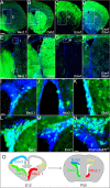
Figure 1.
Figure 1.
All regions of the telencephalic neuroepithelium contribute to the adult SVZ. Coronal sections (30 μm) of adult (P50) mouse brains were stained with rabbit anti-GFP (green) and Hoechst 33258 to visualize cell nuclei (blue). GFP-positive cells were detected by immunolabeling in sections from R26-GFP reporters carrying Nkx2.1-Cre (A, E), Gsh2-Cre (B, F), Emx1-Cre (C, G), or Emx1/Dbx1-Cre (D, H) transgenes. Each region of the embryonic neuroepithelium contributed preferentially to distinct regions of the adult SVZ. Images (I–M) are higher-magnification confocal images of the areas boxed in E–H. I, J, Nkx2.1-Cre/R26-GFP brain. Groups of GFP-positive cells were occasionally detected at the ventral edge of the dorsolateral corner of the SVZ (I), as well as the ventral region of the lateral wall (J). K, L, Gsh2-Cre/R26-GFP brain. Many GFP-positive cells were found in the lateral wall (K) and the dorsolateral corner (L) of the SVZ. M, Emx1-Cre/R26-GFP brain. Significant numbers of GFP-labeled cells are present, mainly in the dorsal half of the dorsolateral corner of the SVZ. N, Adult (P50) Emx1-CreERT2/R26-YFP brain, after tamoxifen induction at E10.5. The distribution of GFP-labeled cells was found to be similar to the Emx1-Cre/R26-GFP animals (i.e., in the dorsal part of the dorsolateral corner of the SVZ). O, Schematic depicting the different embryonic neuroepithelial domains targeted by our Cre mice and their relative contribution to generating the adult SVZ. Scale bars: A–D, 1 mm; E–H, 0.5 mm; I–N, 25 μm.
Figure 2.
Figure 2.
The embryonic striatum and cortex both contribute proliferative cells to the adult SVZ. BrdU was administered to adult R26-GFP reporter mice carrying Nkx2.1-Cre (A, B), Gsh2-Cre (C, D), or Emx1-Cre (E, F) transgenes, and the brains were analyzed 24 h later by immunolabeling with anti-BrdU (red) and anti-GFP (green) and confocal fluorescence microscopy. BrdU/GFP double-positive cells (yellow) were present in all animals, the greatest proportion being found in Gsh2-Cre/R26-GFP mice. The inset high-magnification images are of the cells indicated by arrows (B, D, F). All images are single confocal scans. Scale bars: A–C, 0.5 mm; D–F, 50 μm.
Figure 3.
Figure 3.
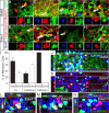
All regions of the embryonic neuroepithelium give rise to multipotent, self-renewing stem cells in the adult SVZ. A, Neurosphere cultures were generated from the SVZ of adult R26-GFP reporter mice carrying Nkx2.1-Cre, Gsh2-Cre, or Emx1-Cre transgenes, or both Emx1- and Dbx1-Cre together. Individual neurospheres were either uniformly GFP positive or negative, consistent with each being derived from a single cell in the starting culture. B, The proportions of GFP-positive neurospheres are shown graphically (mean ± SD; three independent cultures from three adult mice of each genotype). C, Neurospheres were replated under differentiation-inducing conditions for 5 d before being fixed and triple-immunolabeled with anti-GFP (green), anti-βIII-tubulin (red, to label neurons), and monoclonal O4 (magenta, to label oligodendrocyte lineage cells), and then counterstained with 4′,6′-diamidino-2-phenylindole (DAPI) nuclear stain (blue). The example shown in C depicts a GFP-positive and a GFP-negative neurosphere from an Emx1-Cre/R26-GFP culture differentiating side by side, illustrating the generation of neurons and oligodendrocytes from stem cells of differing embryonic origins. D, The proportions of GFP-positive neurospheres of each genotype that generated βIII-tubulin-positive neurons are presented graphically. Nkx2.1-derived neurospheres were significantly less neurogenic than the others (p < 0.05). All neurospheres regardless of genotype generated GFAP-positive astrocytes (data not shown). E, SVZ cells were dissected free of surrounding tissue, dissociated, and cultured in a semisolid matrix with mitogens for 3 weeks (see Materials and Methods). Long-term self-renewing stem cells gave rise to large colonies that distinguished them from transit-amplifying precursors, which have less proliferative potential. Individual colonies were either GFP positive (E, F) or GFP negative (G, H) depending on the origin of the founder stem cell. The examples shown are from an adult Emx1-Cre/R26-GFP culture. I, The proportions of GFP-positive stem cell colonies of each genotype are shown graphically. Scale bars: A, 100 μm; C, 25 μm; E–H, 0.5 mm.
Figure 4.
Figure 4.
Cortex- and LGE-derived SVZ stem cells generate new neuroblasts in vivo. Coronal sections through the forebrain of R26-GFP reporters carrying either the Gsh2-Cre (A, D), Emx1-Cre (B, E), or Emx1-CreERT2 (C, F) transgene were double immunolabeled for GFP (green) together with either GFAP (red; to identify multipotent stem cells) (A–C) or PSA-NCAM (red; to identify neuroblasts) (D–F). All sections were counterstained with Hoechst nuclear dye (blue). Images show the dorsolateral corner of the SVZ (compressed confocal series with orthogonal views taken at the level indicated by the dashed line). GFAP-positive stem cells and PSA-NCAM-positive neuroblasts descend from both the embryonic LGE and cortex (single confocal scan inset). Scale bar, 30 μm.
Figure 5.
Figure 5.
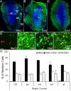
LGE- and cortex-derived stem cells give rise to new cells in the adult olfactory bulb. Thirty micrometer coronal sections through the olfactory bulb of adult R26R-GFP reporter mice carrying Nkx2.1-Cre, Gsh2-Cre, or Emx1-Cre transgenes were immunolabeled for GFP (A–C). To identify newly generated cells, we injected BrdU at P50 and analyzed the olfactory bulb 30 d later (P80) for the presence of (GFP, BrdU) double-positive cells. Colabeled cells were rarely detected in Nkx2.1-Cre/R26-GFP olfactory bulbs (D, H). However, many (GFP, BrdU) double-positive cells were present in Gsh2-Cre/R26-GFP, Emx1-Cre/R26-GFP, and Emx1/Dbx1-Cre/R26-GFP olfactory bulbs (E–G, arrows). The proportions of newly born adult cells were quantified according to genotype for the entire olfactory bulb (OB) and separately for the granule cell layer (gcl), mitral cell layer (mcl), external plexiform layer (epl), and glomerular layer (gl) (see A). There is laminar variation in the relative proportions of Gsh2- and Emx1-derived adult-born cells. For experimental details, see Materials and Methods, Immunocytochemistry. Scale bars: A–C, 200 μm; D–G, 20 μm. Error bars indicate SD. osn, Olfactory sensory neurons.
Figure 6.
Figure 6.
Locations and connections of interneurons in the olfactory bulb. The diagram represents a coronal section through an adult mouse olfactory bulb. New neuroblasts (Dcx positive) enter the bulb from the RMS, migrate radially and differentiate as interneurons in (1) the granule cell layer (GCL), (2) the mitral cell layer (MCL), (3) the EPL, and (4) the glomerular layer (GL). Immunolabeling for calretinin (Crt), calbindin (Cb), tyrosine hydroxylase (TH), or parvalbumin (PV) identifies subpopulations of olfactory interneurons distributed throughout the bulb as shown. The majority of granule neurons (G) are not identified by any of these markers. The MCL also contains mitral projection neurons (M), which are not replaced during adulthood from RMS neuroblasts. Olfactory sensory neurons (O) in the olfactory epithelium in the nasal cavity are also not replaced by RMS neuroblasts. Adapted from Lledo et al. (2006).
Figure 7.
Figure 7.
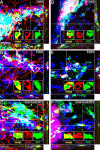
LGE- and cortex-derived SVZ stem cells generate different subpopulations of olfactory interneurons in the adult. BrdU was administered to adult (7 weeks of age) R26-GFP reporter mice carrying either Gsh2-Cre or Emx1-Cre transgenes. Four weeks later (∼P80), coronal sections of olfactory bulbs were triple-immunolabeled for BrdU (blue), GFP (green), and parvalbumin (A, B), tyrosine hydroxylase (TH) (C, D), calretinin (Crt) (E, F), or calbindin (Cb) (G, H) (red). Cells indicated by arrows in the compressed confocal series are shown at high magnification in the insets (single confocal scans). The proportions of adult-born interneurons of each genotype are presented graphically in I. Error bars indicate SD. Gsh2- and Emx1-derived stem cells generate different proportions of TH-, Crt-, and Cb-positive neurons, the most striking example being Cb-positive neurons, which were exclusively Gsh2 (LGE) derived. The vast majority of Cb-positive neurons (red) coexpress GFP (green) in olfactory bulb sections from Gsh2-Cre/R26-GFP (J) but not Emx1-Cre/R26-GFP mice (K), demonstrating that production of Cb-positive interneurons is the preserve of striatum-derived precursor/stem cells throughout development as well as in the adult. L–N, Tamoxifen was administered to Emx1-CreERT2/R26-GFP transgenic mice at E10.5 and the progeny of these cells were traced into adulthood (P50). The olfactory bulbs were immunolabeled to detect GFP (green) and either TH (L), Crt (M), or Cb (N) (red). All nuclei were counterstained with Hoechst 33258 (blue). These data demonstrate that cells derived from the embryonic cortex make a significant contribution to the genesis of TH-positive and Crt-positive interneurons, but fail to generate Cb-positive interneurons. Compressed confocal Z-series are shown with orthogonal views taken at the levels indicated by the dashed lines. For experimental details, see Materials and Methods, Immunocytochemistry. Scale bars: A–H, 20 μm; J–N, 30 μm.
Figure 8.
Figure 8.
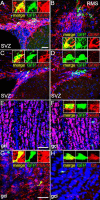
A population of adult SVZ stem cells expresses Emx1 into adulthood. Tamoxifen was administered to young adult (7 weeks of age) Emx1-CreERT2/R26-YFP transgenic mice. After 1 week (A, B) or 6 weeks (C, D), brain sections were double immunolabeled for YFP (green) together with GFAP (red) to label stem cells in the SVZ (A, C, D) or proximal RMS (B). Sections were counterlabeled with Hoechst nuclear stain (blue). (YFP, GFAP) Double-positive stem cells were present at both 1 and 6 weeks after tamoxifen. YFP-single-positive presumptive neuroblasts were also present at 6 weeks (D). No YFP-labeled neurons (NeuN-positive) were present in the olfactory bulb at 1 week after tamoxifen (E), but small numbers were found at 6 weeks (F). At 6 weeks after tamoxifen, (YFP, PSA-NCAM) double-positive neuroblasts and (YFP, calretinin) double-positive interneurons were also present (G, H). High-magnification images of representative cells are shown as insets (single confocal scans). These data are consistent with the interpretation that Emx1-positive stem cells in the SVZ generate neuroblasts that migrate via the RMS into the olfactory bulb and differentiate as olfactory interneurons. Scale bars, 50 μm.

Similar articles

Cited by

References

    1. Alvarez-Buylla A, Lim DA. For the long run: maintaining germinal niches in the adult brain. Neuron. 2004;41:683–686. - PubMed
    1. Anthony TE, Klein C, Fishell G, Heintz N. Radial glia serve as neuronal progenitors in all regions of the central nervous system. Neuron. 2004;41:881–890. - PubMed
    1. Bedard A, Parent A. Evidence of newly generated neurons in the human olfactory bulb. Brain Res Dev Brain Res. 2004;151:159–168. - PubMed
    1. Bull ND, Bartlett PF. The adult mouse hippocampal progenitor is neurogenic but not a stem cell. J Neurosci. 2005;25:10815–10821. - PMC - PubMed
    1. Cao BJ, Li Y. Reduced anxiety- and depression-like behaviors in Emx1 homozygous mutant mice. Brain Res. 2002;937:32–40. - PubMed

Publication types