Comparative Study
. 2007 Aug 1;27(31):8334-43.
doi: 10.1523/JNEUROSCI.2155-07.2007.

NMDA di-heteromeric receptor populations and associated proteins in rat hippocampus

Affiliations
Comparative Study

NMDA di-heteromeric receptor populations and associated proteins in rat hippocampus

Rana A Al-Hallaq et al. J Neurosci. .

Abstract

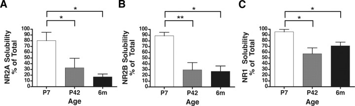
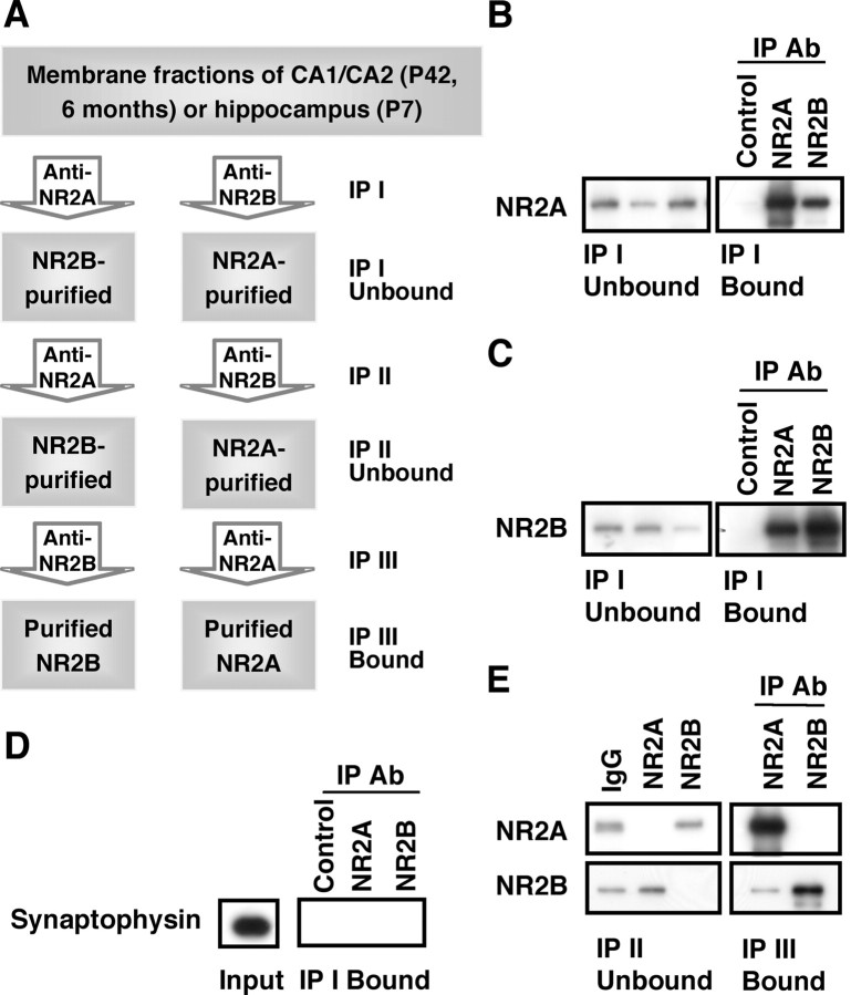
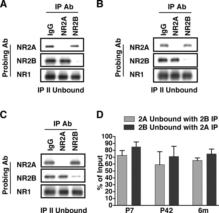
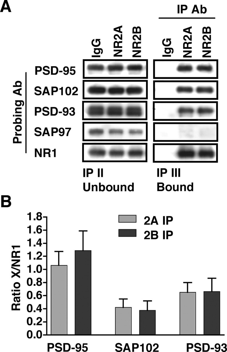
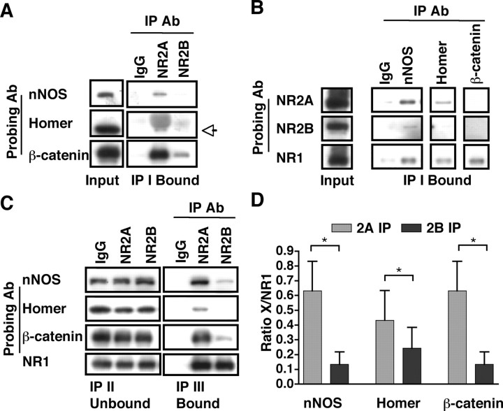
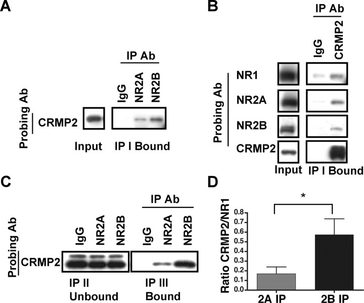
Subunit composition of NMDA receptors (NMDARs) determines a range of physiological properties, downstream signaling effects, and binding partners. Differential localization of NR2A- or NR2B-containing NMDARs within the neuron and subunit-specific protein associations may explain differences in NR2A and NR2B contributions to synaptic plasticity and excitotoxic cell death. This question is complicated by the existence of tri-heteromeric complexes (NR1/NR2A/NR2B). To date, no quantitative biochemical determinations have been made of the relative abundance of different NMDAR populations in intact hippocampus, the region extensively correlated with NMDAR-dependent long-term potentiation. We investigated subunit composition and subunit-specific interactions in CA1/CA2 of rat hippocampus. Using sequential immunoprecipitations to deplete either NR2B or NR2A, di-heteromeric NR1/NR2A and NR1/NR2B receptor populations were isolated from postnatal day 7 (P7) hippocampus and P42 and 6-month-old CA1/CA2. Quantitative Western blot analysis revealed that 60-70% of NR2A and 70-85% of NR2B subunits were associated in NR1/NR2A or NR1/NR2B di-heteromeric complexes. Isolated di-heteromeric receptor fractions were used to examine NR2A- or NR2B-specific interactions with synapse-associated proteins. Our results indicate that NR2A- or NR2B-containing NMDARs associate similarly with postsynaptic density-95 (PSD-95), synapse-associated protein 102, and PSD-93 at P42. However, NR2A-containing receptors coimmunoprecipitated a greater proportion of the synaptic proteins neuronal nitric oxide synthase, Homer, and beta-catenin. Finally, mass spectrometry analysis of isolated di-heteromeric receptors identified a novel NMDAR interactor, collapsin response mediator protein 2, which preferentially associates with NR2B-containing di-heteromeric NMDARs. In summary, in rat hippocampus, NR2A and NR2B exist primarily in di-heteromeric complexes that interact similarly with PSD-95-related proteins but are associated with different protein complexes.


Figures

Figure 1.
Figure 1.
NR1, NR2A, and NR2B detergent solubility decreases with developmental age. Rat whole hippocampus (P7) or CA1/CA2 [P42 and 6 months (6m)] membrane fractions were solubilized with 1% DOC, resolved by SDS-PAGE, and subjected to densitometric analysis. The solubilized fraction is presented as a percentage of the original homogenate for NR2A (A), NR2B (B), and NR1 (C). NMDAR subunit solubility is significantly decreased at P42 and 6 months relative to P7 for the subunits tested. Data are representative of three independent experiments performed on different hippocampal or CA1/CA2 preparations. Values are shown as mean ± SEM. *p < 0.05; **p < 0.01.
Figure 2.
Figure 2.
NR2A and NR2B di-heteromeric receptor populations are isolated by serial immunoprecipitations. A, A flowchart of the experimental procedure outlining the serial immunoprecipitations used to examine di-heteromeric receptors and their associated proteins. The IP II Unbound fraction corresponds to the NR2A- or NR2B-associated di-heteromeric populations. B, C, P42 hippocampal membrane fractions were immunoprecipitated by normal rabbit serum, anti-NR2A, or anti-NR2B antibodies (Ab), and the IP I Unbound and IP I Bound fractions were immunoblotted with antibodies to NR2A (B) or NR2B (C). The anti-NR2A and -NR2B antibodies efficiently immunoprecipitate their respective antigen as well as smaller amounts of the other subunit. D, In similar experiments, NR2A- or NR2B-bound fractions were subjected to immunoblotting with an antibody to synaptophysin. A strong signal was detected in the input, but no signal was detected in the IP I Bound fractions, suggesting that the NR2A and NR2B immunoprecipitations are specific for an NMDAR protein complex. E, Representative blots after double and triple immunoprecipitations with normal rabbit serum, anti-NR2A, or anti-NR2B antibodies. The IP II Unbound fractions verify that the NR2A-depleted (lane 2) or NR2B-depleted (lane 3) fractions do not contain detectable level of NR2A or NR2B, respectively. The IP III Bound fractions indicate that the NR2B-depleted fraction (lane 1) contains NR2A but a low level of NR2B, and the NR2A-depleted fraction (lane 2) contains NR2B but no detectable levels of NR2A, confirming efficient isolation of NR2A- or NR2B-containing di-heteromeric receptor populations. For all experiments, 14 μl of the input, unbound, and bound fractions were resolved by SDS-PAGE and probed. Because the bound fractions were resuspended in 0.1 vol of the original input, the bound sample is 10 times more concentrated than the input or unbound fractions.
Figure 3.
Figure 3.
NR2A and NR2B are in di-heteromeric NR1/NR2A or NR1/NR2B or in tri-heteromeric NR1/NR2A/NR2B complexes throughout development. P42 CA1/CA2 (A, D), P7 hippocampus (B, D), and 6-month-old CA1/CA2 (C, D) were subjected to double immunoprecipitations with anti-NR2A or anti-NR2B antibodies (Ab). A–C, Representative immunoblots of the IP II Unbound fractions probed with anti-NR2A, -NR2B, or -NR1 antibodies demonstrating that the majority of NR2A (lane 2) or NR2B (lane 3) has been depleted (compare with normal rabbit serum, lane 1). D, Densitometric analysis of P7 hippocampus, P42 CA1/CA2, and 6-month-old (6m) CA1/CA2 of the unbound fraction normalized to the input. Data are representative of three independent experiments performed on different hippocampal or CA1/CA2 preparations. Values are shown as mean ± SEM.
Figure 4.
Figure 4.
PSD-95, SAP102, and PSD-93 associate to a similar extent with the NR1/NR2A or NR1/NR2B di-heteromeric receptors in P42 CA1/CA2. Immunoprecipitations with anti-NR2A and anti-NR2B antibodies from isolated di-heteromeric receptor fractions (NR1/NR2A and NR1/NR2B) were analyzed for coassociation with the PSD-95 family of proteins. A, Representative immunoblots of the IP II Unbound and IP III Bound fractions after triple immunoprecipitations and immunoblotting with anti-PSD-95, -SAP102, -PSD-93, -SAP97, and -NR1 antibodies (Ab). With the exception of SAP97, NR2A- and NR2B-bound fractions coimmunoprecipitate similar levels of the proteins investigated. B, Densitometric analysis of the levels of PSD-95, SAP102, or PSD-93 coimmunoprecipitated with NR2A- and NR2B-containing di-heteromeric receptors. All data are normalized to the level of NMDAR immunoprecipitated, as measured by NR1 coimmunoprecipitations, and no significant differences are found between NR2A- or NR2B-immunoprecipitated fractions for any of the proteins. Data are representative of eight (PSD-95 and SAP102) or four (PSD-93) independent experiments performed on different CA1/CA2 preparations. Values are shown as mean ± SEM.
Figure 5.
Figure 5.
The synaptic proteins nNOS, Homer, and β-catenin associate to a greater extent with the di-heteromeric NR1/NR2A receptors than with NR1/NR2B receptors. A, Representative blots are shown of single immunoprecipitation experiments with anti-NR2A or -NR2B antibodies (Ab), probed with anti-nNOS, Homer, and β-catenin antibodies. Both NR2A and NR2B coimmunoprecipitate the synapse-associated proteins; however, the interaction with NR2A appears to be stronger. The Homer band at 47 kDa (noted by the arrow) may be distinguished from the higher-molecular-weight heavy chain IgG bands. B, Single immunoprecipitation experiments with anti-nNOS, Homer, and β-catenin antibodies probed with anti-NR2A, NR2B, and NR1. NR1 is coimmunoprecipitated with nNOS, Homer, and β-catenin, NR2A coimmunoprecipitates with nNOS and Homer, and NR2B co-associates with nNOS, with no detectable signal in anti-Homer or anti-β-catenin immunoprecipitations. C, D, Immunoprecipitations with anti-NR2A and anti-NR2B antibodies from isolated di-heteromeric receptor fractions (NR1/NR2A and NR1/NR2B) examined above were analyzed for coassociation of nNOS, Homer, and β-catenin. C, Representative blots of the IP II Unbound and IP III Bound fractions after triple immunoprecipitations and immunoblotting with anti-nNOS, -Homer, and -β-catenin antibodies. NR1 blots are identical to those in Figure 4. Densitometric analysis (D) of nNOS, Homer, or β-catenin associated with NR2A and NR2B, normalized to the level of NMDAR immunoprecipitated as measured by coimmunoprecipitated NR1. Data are representative of four (nNOS and β-catenin) or seven (Homer) independent experiments performed on different hippocampal preparations. Values are shown as mean ± SEM. *p < 0.05.
Figure 6.
Figure 6.
CRMP2 interacts preferentially with the di-heteromeric NR1/NR2B receptors. A, A representative blot of the input and IP I Bound fractions after immunoprecipitations with anti-NR2A and -NR2B antibodies (Ab). Immunoblot analysis with an anti-CRMP2 antibody demonstrated that CRMP2 coimmunoprecipitates with NR2A and NR2B from P42 hippocampus. B, Representative blots of the input and IP I Bound fractions after immunoprecipitations with an anti-CRMP2 antibody. Immunoblot analysis with anti-NR1, -NR2A, or -NR2B antibodies demonstrated that CRMP2 coimmunoprecipitates the NMDAR subunits from P42 hippocampus. C, Immunoprecipitations with anti-NR2A and anti-NR2B antibodies from isolated di-heteromeric receptor fractions were analyzed for coassociation of CRMP2. A representative blot of the IP II Unbound and IP III Bound fractions after immunoblotting with anti-CRMP2 antibodies. CRMP2 associates to a greater extent with the NR2B subunit. D, Densitometric analysis of CRMP2 coimmunoprecipitated with NR2A and NR2B, normalized to the level of NMDAR immunoprecipitated as measured by coimmunoprecipitated NR1. Data are representative of six independent experiments performed on different CA1/CA2 preparations. Values are shown as mean ± SEM. *p < 0.05.

Similar articles

Cited by

References

    1. Al-Hallaq RA, Jarabek BR, Fu Z, Vicini S, Wolfe BB, Yasuda RP. Association of NR3A with the N-methyl-d-aspartate receptor NR1 and NR2 subunits. Mol Pharmacol. 2002;62:1119–1127. - PubMed
    1. Allison DW, Gelfand VI, Spector I, Craig AM. Role of actin in anchoring postsynaptic receptors in cultured hippocampal neurons: differential attachment of NMDA versus AMPA receptors. J Neurosci. 1998;18:2423–2436. - PMC - PubMed
    1. Allison DW, Chervin AS, Gelfand VI, Craig AM. Postsynaptic scaffolds of excitatory and inhibitory synapses in hippocampal neurons: maintenance of core components independent of actin filaments and microtubules. J Neurosci. 2000;20:4545–4554. - PMC - PubMed
    1. Arimura N, Menager C, Kawano Y, Yoshimura T, Kawabata S, Hattori A, Fukata Y, Amano M, Goshima Y, Inagaki M, Morone N, Usukura J, Kaibuchi K. Phosphorylation by Rho kinase regulates CRMP-2 activity in growth cones. Mol Cell Biol. 2005;25:9973–9984. - PMC - PubMed
    1. Besshoh S, Bawa D, Teves L, Wallace MC, Gurd JW. Increased phosphorylation and redistribution of NMDA receptors between synaptic lipid rafts and post-synaptic densities following transient global ischemia in the rat brain. J Neurochem. 2005;93:186–194. - PubMed

Publication types