Comparative Study
. 2007 Aug 1;27(31):8378-86.
doi: 10.1523/JNEUROSCI.0872-07.2007.

RhoA regulates dendrite branching in hippocampal neurons by decreasing cypin protein levels

Affiliations
Comparative Study

RhoA regulates dendrite branching in hippocampal neurons by decreasing cypin protein levels

Hongxin Chen et al. J Neurosci. .

Abstract

The way a dendrite is patterned determines how a neuron will receive information. The Rho GTPases have been reported to play increasingly well defined roles in determining dendritic branch and spine development and morphology. Much is known about how these small GTPases regulate the actin cytoskeleton; however, very little is known about how they alter the microtubule cytoskeleton. Our laboratory previously cloned and characterized cypin, a guanine deaminase that increases dendrite number by binding to tubulin heterodimers and promoting microtubule assembly. In the present study, we show that cypin and RhoA are part of a common pathway that regulates dendrite number. Inhibition of Rho kinase activity does not affect cypin-mediated dendrite branching. Furthermore, cypin does not affect the activity of RhoA, as measured by GTP binding to RhoA. In fact, activated RhoA acts to inhibit cypin protein expression and, by doing so, decreases dendrite number. In addition, this decrease in cypin protein occurs via a translation-dependent mechanism. Together, our data suggest that cypin acts downstream of the small GTPase RhoA to regulate dendrite branching in hippocampal neurons, providing a novel mechanism for RhoA action on microtubule dynamics.


Figures

Figure 1.
Figure 1.
RhoA and cypin are part of a signaling pathway that regulates dendrite number. RhoCA and RhoDN were coexpressed with either GFP or GFP–cypin in hippocampal neurons from DIV 10–12. Neurons were fixed and immunostained for dendrite counting at DIV 12. A, GFP images of representative neurons. Scale bar, 10 μm. B, Average number of primary and secondary dendrites in transfected neurons. n values are as follows: GFP, n = 35; cypin, n = 24; GFP/RhoCA, n = 54; cypin/RhoCA, n = 27; GFP/RhoDN, n = 14; cypin/RhoDN, n = 19. *p < 0.05, **p < 0.01, ***p < 0.001 by Kruskal–Wallis test followed by Dunn's multiple comparisons test compared with GFP. C, Sholl analysis of transfected neurons. Overexpression of cypin (red circles) significantly increases number of intersections at 20–35 and 45 μm from the soma compared with overexpression of GFP (black squares; p < 0.01 at 25 and 30 μm, p < 0.05 at other points). Left, Overexpression of RhoCA (green triangles) dramatically decreases number of intersections at 5–40 μm from soma compared with overexpression of GFP (black squares; p < 0.05 at 40 μm, p < 0.01 at 20–30 μm, p < 0.001 at other points). Expression of RhoCA blocks cypin-promoted increases in number of intersections, resulting in baseline (GFP) number of intersections. p values were determined by Kruskal–Wallis test followed by Dunn's multiple comparisons test compared with GFP. Right, Coexpression RhoDN with GFP or cypin does not affect dendritic arbor complexity. Error bars indicate SEM.
Figure 2.
Figure 2.
Overexpression of cypin does not affect RhoA activity in Neuro-2a cells. Neuro-2a cells were transfected with cDNA encoding GFP, cypin, or cypin mutants lacking guanine deaminase activity. A, Twenty-four hours after transfection, activated RhoA was isolated from cell lysates using rhotekin–GST coupled to glutathione-Sepharose beads. The eluates were subjected to Western blotting using an antibody to RhoA. Left, Representative blot is shown from three experiments. Right, Bands were quantitated as described in Materials and Methods. There is no significant change in the amount of RhoA activation when cypin or cypin mutants are expressed when compared with when GFP is expressed. (n = 3). B, A colorimetric guanine deaminase activity assay was performed on Neuro-2a cells expressing GFP, cypin, or cypin mutants to confirm that the cypin mutants do not retain guanine deaminase activity (n = 3). Error bars indicate SEM.
Figure 3.
Figure 3.
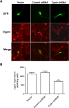
shRNA knocks down cypin protein expression. Hippocampal neurons were transfected with vector, control shRNA, or cypin shRNA at DIV 10. Neurons were fixed and immunostained for GFP and cypin at DIV 12. A, Representative GFP fluorescence images of neurons transfected with cDNAs encoding the stated constructs. Arrows in cypin panels (middle row) indicate neurons transfected with the stated cDNAs. Scale bar, 10 μm. B, After the cells were fixed and immunostained for cypin at DIV 12, cypin immunoreactivity was measured using the ImagePro analysis program. n values are as follows: vector, n = 45; control shRNA, n = 34; cypin shRNA, n = 49. **p < 0.01 by Kruskal–Wallis test followed by Dunn's multiple comparisons test compared with vector. Error bars indicate SEM.
Figure 4.
Figure 4.
A silent mutant of cypin rescues the decreased dendrite number resulting from knockdown of endogenous cypin protein. Hippocampal neurons were cotransfected at DIV 10 with cypin shRNA and cDNA encoding smCypin in the region targeted by our shRNA. Dendrites were counted directly after fixation at DIV 12. A, Sequences of smCypin, wild-type cypin (wtCypin), and the shRNA target (cypin shRNA). smCypin contains four mutated bases compared with wild-type cypin. B, GFP images fluorescence of representative neurons. Scale bar, 10 μm. C, Average number of primary and secondary dendrites in transfected neurons. n values are as follows: vector, n = 30; cypin shRNA, n = 41; cypin shRNA plus smCypin, n = 28. ***p < 0.001 by Kruskal–Wallis test followed by Dunn's multiple comparisons test compared with vector. Error bars indicate SEM.
Figure 5.
Figure 5.
Knockdown of cypin protein expression eliminates the effects of RhoDN on dendrite branching. Hippocampal neurons were transfected with cDNAs encoding the stated constructs at DIV 10. Neurons were fixed and immunostained for dendrite counting at DIV 12. A, GFP images of representative neurons. Scale bar, 10 μm. B, Average number of primary and secondary dendrites in transfected neurons. n values are as follows: vector, n = 17; control shRNA, n = 21; cypin shRNA, n = 33; RhoCA plus vector, n = 20; RhoCA plus control shRNA, n = 18; RhoCA plus cypin shRNA, n = 25; RhoDN plus vector, n = 28; RhoDN plus control shRNA, n = 32; RhoDN plus cypin shRNA, n = 31. *p < 0.05, **p < 0.01 and ##p < 0.01, ###p < 0.001 by Kruskal–Wallis test followed by Dunn's multiple comparisons test compared with untreated vector unless otherwise indicated. Error bars indicate SEM.
Figure 6.
Figure 6.
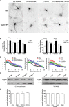
Treatment of neurons with C3 transferase, but not Y27632, blocks cypin-promoted increases in dendrite number without affecting cypin protein expression. Hippocampal neurons were transfected with constructs encoding GFP or cypin–GFP at DIV 10, treated with the appropriate drug at DIV 11 for 24 h, and processed for immunostaining at DIV 12. A, GFP images of representative neurons. Scale bar, 10 μm. B, Average number of primary and secondary dendrites in transfected neurons. n values are as follows: in GFP group: none, n = 35; C3, n = 22; Y27632, n = 20; both, n = 18; and in cypin group: none, n = 24; C3, n = 15; Y27632, n = 15; both, n = 13. #p < 0.05, ##p < 0.01, ###p < 0.001 by ANOVA followed by Bonferroni's multiple comparisons test. **p < 0.01 by ANOVA followed by Dunnett's multiple comparisons test compared with GFP. C, Left, Sholl analysis of hippocampal neurons. Overexpression of cypin significantly increases number of intersections in the range 20–55 μm compared with expression of GFP (red circles; p < 0.01 at 30–45 μm, p < 0.05 at other points). Treatment with C3 transferase does not change dendritic arbor complexity, even combined with expression of cypin (blue x). Middle, Neurons overexpressing cypin and treated with Y27632 (blue x) show a significant increase in number of intersections at 5–75 μm from the soma (except at 10 μm; p < 0.05 at 15, 25–35, 60, and 75 μm, p < 0.01 at other points). Neurons treated with Y27632 (green triangles) alone show increased number of intersections at 35–75 μm from the soma (p < 0.05 at 35 and 75 μm, p < 0.01 at other points). p values were determined by Kruskal–Wallis test followed by Dunn's multiple comparisons test compared with GFP. Right, Neurons overexpressing cypin (blue x) or GFP (green triangles) and treated with both inhibitors show no significant change in number of intersections from that of control (GFP). D, Hippocampal neurons were treated with C3 transferase or Y27632 at indicated concentrations at DIV 11 for 24 h. Representative Western blot of three individual experiments is shown. E, Quantitative analysis shows that treatments do not change is cypin protein expression (p > 0.05 by ANOVA). Error bars indicate SEM.
Figure 7.
Figure 7.
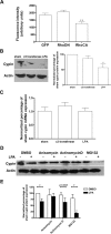
Activation of RhoA decreases cypin protein expression via a translation-dependent mechanism. A, Cultured hippocampal neurons were transfected with cDNAs encoding RhoCA or RhoDN at DIV 10. Neurons were fixed and immunostained at DIV 12, and cypin levels were quantitated using ImagePro software. n values are as follows: GFP, n = 30; RhoCA, n = 36; RhoDN, n = 35. **p < 0.01 by ANOVA followed by Dunnett's multiple comparison test compared with GFP. B, Hippocampal neurons were treated with 100 ng/ml C3 transferase at DIV 11 for 24 h or 1 μg/ml LPA at DIV 12 for 15 min. Quantitation of Western blots shows that there is significant change in cypin protein expression with LPA but not C3 transferase treatment (n = 3). *p < 0.05 by ANOVA followed by Dunnett's multiple comparison test compared with GFP. C, Semiquantitative real-time reverse transcription-PCR shows there is no significant change in cypin mRNA levels with these treatments. D, At DIV 12, hippocampal neurons were preincubated with 40 μm anisomycin, 5 μm actinomycin D, or 50 μm MG132 for 30 min. The neurons were then treated with LPA at 1 μg/ml for 15 min. A representative blot is shown from four experiments. E, Quantitation of Western blots shows that anisomycin, but not actinomycin D or MG132, blocks the LPA-mediated decrease in cypin protein expression. **p < 0.01 by ANOVA followed by Dunnett's multiple comparison test compared with DMSO; #p < 0.05 by paired t test (n = 4). Error bars indicate SEM.
Figure 8.
Figure 8.
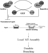
Model for RhoA-regulated dendrite branching. Activation of RhoA decreases cypin protein expression and in turn inhibits local microtubule (MT) assembly and decreases dendrite branching. Inhibition of RhoA will allow cypin protein levels to remain at a constitutive state and disallow the inhibitory effect of activated RhoA on cypin translation and dendrite branching.

Similar articles

Cited by

References

    1. Ahnert-Hilger G, Holtje M, Grosse G, Pickert G, Mucke C, Nixdorf-Bergweiler B, Boquet P, Hofmann F, Just I. Differential effects of Rho GTPases on axonal and dendritic development in hippocampal neurones. J Neurochem. 2004;90:9–18. - PubMed
    1. Akum BF, Chen M, Gunderson SI, Riefler GM, Scerri-Hansen MM, Firestein BL. Cypin regulates dendrite patterning in hippocampal neurons by promoting microtubule assembly. Nat Neurosci. 2004;7:145–152. - PubMed
    1. Arimura N, Kaibuchi K. Key regulators in neuronal polarity. Neuron. 2005;48:881–884. - PubMed
    1. Baas PW, Karabay A, Qiang L. Microtubules cut and run. Trends Cell Biol. 2005;15:518–524. - PubMed
    1. Bishop AL, Hall A. Rho GTPases and their effector proteins. Biochem J. 2000;348:241–255. - PMC - PubMed

Publication types

LinkOut - more resources