. 2007 Aug 1;21(15):1869-81.
doi: 10.1101/gad.1545707.

p21 transcription is regulated by differential localization of histone H2A.Z

Affiliations

p21 transcription is regulated by differential localization of histone H2A.Z

Nicolas Gévry et al. Genes Dev. .

Abstract

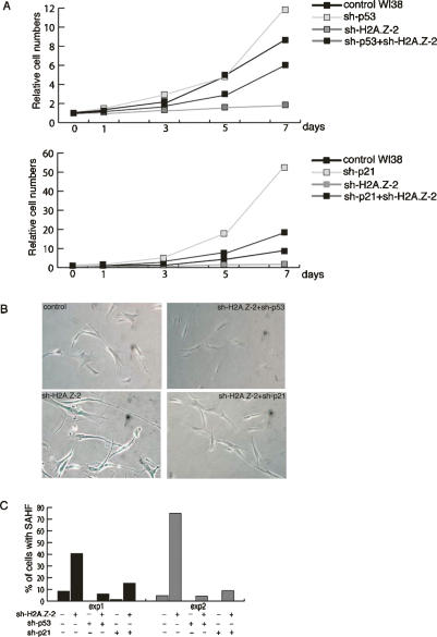
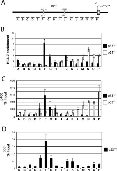
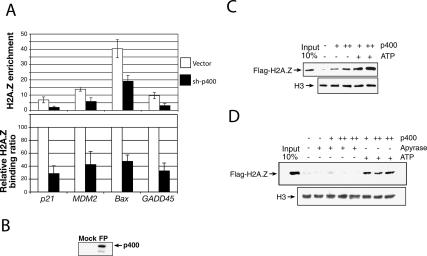
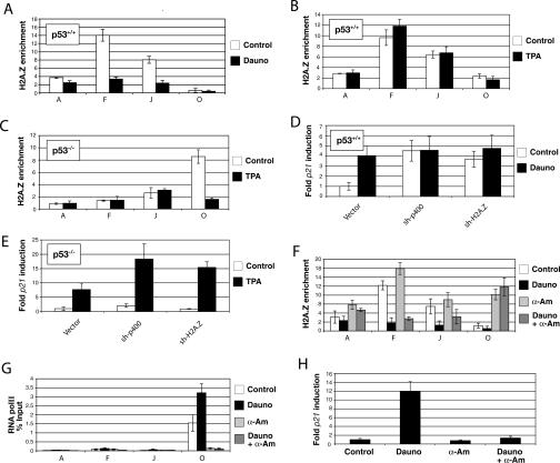
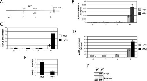
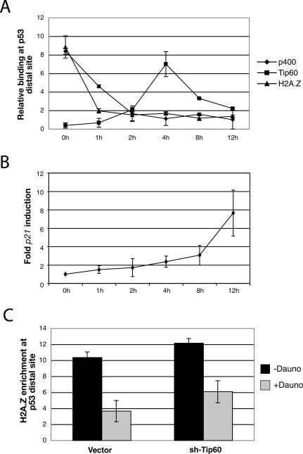
In yeast cells, H2A.Z regulates transcription and is globally associated within a few nucleosomes of the initiator regions of numerous promoters. H2A.Z is deposited at these loci by an ATP-dependent complex, Swr1.com. Here we show that H2A.Z suppresses the p53 --> p21 transcription and senescence responses. Upon DNA damage, H2A.Z is first evicted from the p21 promoter, followed by the recruitment of the Tip60 histone acetyltransferase to activate p21 transcription. p400, a human Swr1 homolog, is required for the localization of H2A.Z, and largely colocalizes with H2A.Z at multiple promoters investigated. Notably, the presence of sequence-specific transcription factors, such as p53 and Myc, provides positioning cues that direct the location of H2A.Z-containing nucleosomes within these promoters. Collectively, this study strongly suggests that certain sequence-specific transcription factors regulate transcription, in part, by preferentially positioning histone variant H2A.Z within chromatin. This H2A.Z-centered process is part of an epigenetic process for modulating gene expression.


Figures

Figure 1.
Figure 1.
H2A.Z represses the basal expression of p21 in p53+/+ cells, and regulates senescence in human fibroblasts. (A) Western blot showing the knockdown of H2A.Z expression by different H2A.Z hairpins. (B, left panel) Expression levels of p21, cyclin D1, and H2Ai in U2OS cells before and after H2A.Z knockdown using shH2A.Z-2. (Right panel) Expression levels of p21 in HCT116 p53+/+ and p53−/− paired cells before and after H2A.Z knockdown using shH2A.Z-2. mRNA expression levels were normalized against expression levels of the 36B4 ribosomal gene, as measured by Q-PCR. (C) FACs analysis of BJ cells following H2A.z depletion. Results from duplicate samples were presented. (D) Cell growth assay performed in BJ cells. (E) Senescent β-gal staining of WI-38 cells following the indicated perturbations. sh-p400 was included as a positive control. (F) DNA staining by DAPI of WI-38 cells following the indicated treatments.
Figure 2.
Figure 2.
Inactivation of p53/p21 rescues H2A.Z depletion-induced senescent phenotypes. (A) Cell growth assay in WI-38 cells following the indicated treatment. (B) Senescent β-gal staining of the WI-38 cells following the indicated treatment. (C) Summary of SAHF in WI-38 cells exposed to the indicated perturbation. The numerical values are listed in Supplementary Table S1. Cells were fixed and counted on days 10 and 12 post-infection in Experiments 1 and 2, respectively.
Figure 3.
Figure 3.
H2A.Z colocalizes with p53 and p400 at the p21 promoter. (A) Diagram of the p21 promoter region and its indigenous p53 response elements, and segments (columns A–P) used for Q-PCR. (B) ChIP analysis of H2A.Z enrichment (percent input of H2A.Z/H2A) at the p21 promoter in U20S (p53+/+) and SAOS2 (p53−/−) cells. (C) ChIP assays for p400 on the p21 promoter in U2OS and SAOS2 cells. (D) ChIP experiments determining the recruitment of p53 at the p21 promoter region in U2OS cells.
Figure 4.
Figure 4.
p400 incorporates H2A.Z into chromatin around selected p53-binding sites. (A) ChIP experiments showing the relative binding of H2A.Z after p400 knockdown at different p53 target genes. The primers used in these experiments correspond to the p53-binding sites for each gene (fragment F [cf. Fig. 3], p21; fragment E, GADD45; fragment F, BAX; fragment E, MDM2). (B) Immunoblot assay with antibody against p400 demonstrating the effect of Flag-specific purification (FP). (C) In vitro exchange of H2A.Z–H2B dimers by p400. Flag-H2A.Z was detected by immunoblotting using an anti-Flag antibody. The H3 immunoblot was used here as a loading control. (D) Similar in vitro exchange of H2A.Z–H2B dimers by p400 but with the addition of apyrase to show dependency of ATP for the reaction.
Figure 5.
Figure 5.
Local eviction of H2A.Z-containing nucleosomes upon induction of the p21 promoter is p53 dependent. (A) H2A.Z enrichment at specific sites within the p21 promoter region (cf. Fig. 4) before and after treatment with 250 μM daunorubicin for 12 h in p53+/+ cells. (B) ChIP analysis of H2A.Z enrichment at the p21 promoter in p53+/+cells treated with 50 ng/mL TPA. (C) ChIP experiments showing the effect of TPA treatment on the localization of H2A.Z in p53−/− cells. The various segments analyzed are also from Figure 4 (D) Relative expression of p21 before and after daunorubicin treatment of p53+/+ cells depleted for H2A.Z and p400. (E) Expression levels of p21 before and after treatment with TPA for 4 h in p53−/− cells depleted in H2A.Z and p400. (F) H2A.Z enrichment at the p21 promoter in p53+/+ cells treated with 250 μM daunorubicin and/or 100 μM α-amanitin for 12 h. (G) ChIP corresponding to the data shown in F, showing binding of RNA polII (8WG16 antibody). (H) p21 expression levels of U2OS cells following various treatments.
Figure 6.
Figure 6.
c-Myc directs H2A.Z recruitment at the p21 TATA region in p53−/− cells. (A) Diagram of the p21 promoter region with the amplicons used for Q-PCR. (B) ChIP assay showing c-Myc binding at the p21 promoter in HaCat-tetMyc cells that overexpress c-Myc under the control of tetracycline. (C) ChIP analysis of H2A.Z enrichment before and after c-Myc overexpression. (D) ChIP analysis of p400 binding before and after c-Myc overexpression. (E) Basal expression level of p21 in HaCat-tetMyc. (F) Immunoblot assay showing c-Myc expression in the HaCat-tetMyc cells cultured for 24 h with or without tetracycline.
Figure 7.
Figure 7.
p400-independent recruitment of Tip60 at the p53-distal site of the p21 promoter. (A) ChIP assay showing the kinetics of H2A.Z, p400, and Tip60 occupancy during p21 promoter activation after daunorubicin treatment in U2OS cells. Q-PCR was performed using primers corresponding to the distal p53 site of the p21 promoter (fragment F). (B) p21 mRNA expression levels after daunorubicin treatment of U2OS cells. (C) ChIP experiment showing H2A.Z enrichment at the distal p53 site after Tip60 knockdown before and after daunorubicin treatment.

References

    1. Adam M., Robert F., Larochelle M., Gaudreau L., Robert F., Larochelle M., Gaudreau L., Larochelle M., Gaudreau L., Gaudreau L. H2A.Z is required for global chromatin integrity and for recruitment of RNA polymerase II under specific conditions. Mol. Cell. Biol. 2001;21:6270–6279. - PMC - PubMed
    1. Allis C.D., Glover C.V., Bowen J.K., Gorovsky M.A., Glover C.V., Bowen J.K., Gorovsky M.A., Bowen J.K., Gorovsky M.A., Gorovsky M.A. Histone variants specific to the transcriptionally active, amitotically dividing macronucleus of the unicellular eucaryote, Tetrahymena thermophila. Cell. 1980;20:609–617. - PubMed
    1. Bakshi R., Mehta A.K., Sharma R., Maiti S., Pasha S., Brahmachari V., Mehta A.K., Sharma R., Maiti S., Pasha S., Brahmachari V., Sharma R., Maiti S., Pasha S., Brahmachari V., Maiti S., Pasha S., Brahmachari V., Pasha S., Brahmachari V., Brahmachari V. Characterization of a human SWI2/SNF2 like protein hINO80: Demonstration of catalytic and DNA binding activity. Biochem. Biophys. Res. Commun. 2006;339:313–320. - PubMed
    1. Berns K., Hijmans E.M., Mullenders J., Brummelkamp T.R., Velds A., Heimerikx M., Kerkhoven R.M., Madiredjo M., Nijkamp W., Weigelt B., Hijmans E.M., Mullenders J., Brummelkamp T.R., Velds A., Heimerikx M., Kerkhoven R.M., Madiredjo M., Nijkamp W., Weigelt B., Mullenders J., Brummelkamp T.R., Velds A., Heimerikx M., Kerkhoven R.M., Madiredjo M., Nijkamp W., Weigelt B., Brummelkamp T.R., Velds A., Heimerikx M., Kerkhoven R.M., Madiredjo M., Nijkamp W., Weigelt B., Velds A., Heimerikx M., Kerkhoven R.M., Madiredjo M., Nijkamp W., Weigelt B., Heimerikx M., Kerkhoven R.M., Madiredjo M., Nijkamp W., Weigelt B., Kerkhoven R.M., Madiredjo M., Nijkamp W., Weigelt B., Madiredjo M., Nijkamp W., Weigelt B., Nijkamp W., Weigelt B., Weigelt B., et al. A large-scale RNAi screen in human cells identifies new components of the p53 pathway. Nature. 2004;428:431–437. - PubMed
    1. Braig M., Lee S., Loddenkemper C., Rudolph C., Peters A.H., Schlegelberger B., Stein H., Dorken B., Jenuwein T., Schmitt C.A., Lee S., Loddenkemper C., Rudolph C., Peters A.H., Schlegelberger B., Stein H., Dorken B., Jenuwein T., Schmitt C.A., Loddenkemper C., Rudolph C., Peters A.H., Schlegelberger B., Stein H., Dorken B., Jenuwein T., Schmitt C.A., Rudolph C., Peters A.H., Schlegelberger B., Stein H., Dorken B., Jenuwein T., Schmitt C.A., Peters A.H., Schlegelberger B., Stein H., Dorken B., Jenuwein T., Schmitt C.A., Schlegelberger B., Stein H., Dorken B., Jenuwein T., Schmitt C.A., Stein H., Dorken B., Jenuwein T., Schmitt C.A., Dorken B., Jenuwein T., Schmitt C.A., Jenuwein T., Schmitt C.A., Schmitt C.A. Oncogene-induced senescence as an initial barrier in lymphoma development. Nature. 2005;436:660–665. - PubMed

Publication types

MeSH terms