. 2007 Oct;18(10):4024-36.
doi: 10.1091/mbc.e07-05-0517. Epub 2007 Aug 1.

Use of the novel Plk1 inhibitor ZK-thiazolidinone to elucidate functions of Plk1 in early and late stages of mitosis

Affiliations

Use of the novel Plk1 inhibitor ZK-thiazolidinone to elucidate functions of Plk1 in early and late stages of mitosis

Anna Santamaria et al. Mol Biol Cell. 2007 Oct.

Abstract

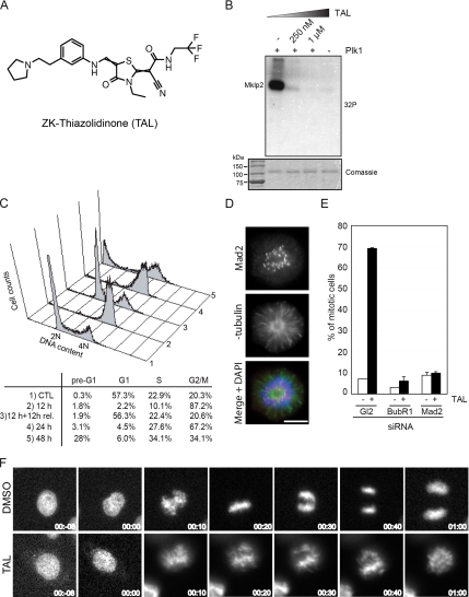
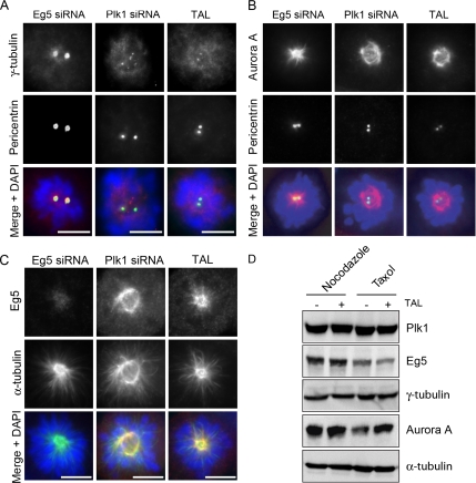
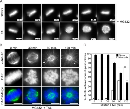
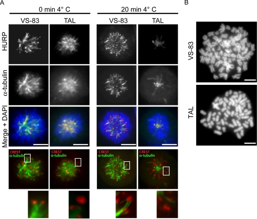
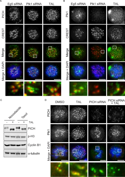
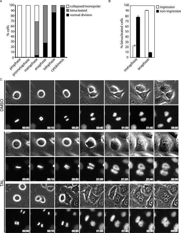
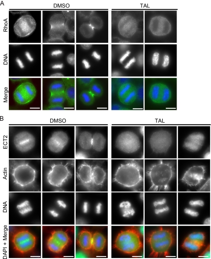
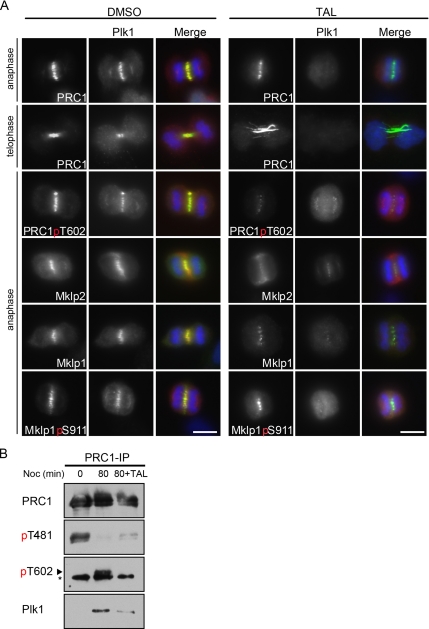
Polo-like kinase 1 (Plk1) is a key regulator of mitotic progression and cell division in eukaryotes. It is highly expressed in tumor cells and considered a potential target for cancer therapy. Here, we report the discovery and application of a novel potent small-molecule inhibitor of mammalian Plk1, ZK-Thiazolidinone (TAL). We have extensively characterized TAL in vitro and addressed TAL specificity within cells by studying Plk1 functions in sister chromatid separation, centrosome maturation, and spindle assembly. Moreover, we have used TAL for a detailed analysis of Plk1 in relation to PICH and PRC1, two prominent interaction partners implicated in spindle assembly checkpoint function and cytokinesis, respectively. Specifically, we show that Plk1, when inactivated by TAL, spreads over the arms of chromosomes, resembling the localization of its binding partner PICH, and that both proteins are mutually dependent on each other for correct localization. Finally, we show that Plk1 activity is essential for cleavage furrow formation and ingression, leading to successful cytokinesis.


Figures

Figure 1.
Figure 1.
TAL inhibits specifically Plk1 and cause a prometaphase-like mitotic arrest. (A) Chemical structure of the ATP-competitive kinase inhibitor, ZK-Thiazolidinone (TAL). (B) Mklp2 was treated with buffer alone (−Plk1) or Plk1 with increasing amounts of TAL and radioactive ATP for 30 min and then analyzed by SDS-PAGE and autoradiography ([32P]). Comassie blue staining showed equal amounts of Mklp2 in all samples. (C) FACS analysis was performed in HeLa S3 cells treated for 24 h with DMSO as a control (1) or for 12 h, 12 h + 12 h release, 24 h, and 48 h with 1 μM TAL (2, 3, 4, and 5, respectively). The percentage of cells with less than 2N (pre-G1), 2N (G1 and S), or 4N (G2/M) content are shown. (D) Mad2-positive staining in a monopolar cell treated with 1 μM TAL. Cells were stained for Mad2 (red), α-tubulin (green), and DNA was stained with DAPI (blue). Scale bar, 10 μm. (E) Mitotic index from cells after transfection with GL2, BubR1, and Mad2 siRNA-oligos for 48 h, treated with either DMSO or 1 μM TAL for the last 12 h. (300 mitotic cells for each combination, n = 2). (F) Selected live-cell images of HeLa cells expressing GFP-tagged Histon-H2B treated with DMSO or 1 μM TAL. Time = 0 min indicates the onset of chromosome condensation.
Figure 2.
Figure 2.
Centrosome maturation is impaired in TAL-treated cells. HeLa S3 cells were transfected with Eg5 or Plk1 siRNA-oligos for 36 h or treated with 1 μM TAL for 12 h and then stained for γ-tubulin, Aurora A, or Eg5 (red; A, B, and C, respectively), pericentrin (A and B), or α-tubulin (C; green), and DNA was stained with DAPI (blue). Scale bar, 10 μm. (D) Lysates from nocodazole- (0.2 μg/mL) or taxol- (1 μg/ml) arrested cells, treated with DMSO or 1 μM TAL were used for Western blot analysis. Membranes were probed for Plk1, Eg5, γ-tubulin, Aurora A, and α-tubulin as loading control.
Figure 3.
Figure 3.
Plk1 activity is required for bipolar spindle maintenance. (A) Cells were arrested with 20 μM VS-83 for 12 h and released into 20 μM MG132 for 2 h to enrich in metaphase cells. Then 1 μM TAL was added and cells were followed by live-cell imaging. Representative stills from live-cell images of HeLa cells expressing GFP-tagged Histone-H2B are shown. Time = 0 represents the time of addition of the inhibitor. (B) HeLa S3 cells were arrested with 25 μM noscapine during 10 h and release into 20 μM MG132. After 2-h MG132 arrest, 1 μM TAL was added, and cells were fixed at indicated times and stained for α-tubulin (green), and DNA was stained with DAPI (blue). Representative images of phenotypes observed at each time point are shown. Scale bar, 10 μm. (C) Quantification of mono- or bipolar spindles in TAL-treated cells; 300 mitotic cells for each data point, n = 2.
Figure 4.
Figure 4.
(A) Plk1 activity is required for K-fiber stabilization and chromatid arm separation. HeLa S3 cells were treated for 12 h with 20 μM VS-83 or 1 μM TAL and before fixation and permeabilization with PTEMF; cells were incubated for 20 min in ice-cold growth medium. Cells were stained for HURP (far red), CREST (red), and α-tubulin (green), and DNA was stained with DAPI (blue). (B) DAPI-stained chromosome spread prepared from HeLa S3 treated for 10 h with 20 μM VS-83 or 1 μM TAL.
Figure 5.
Figure 5.
Plk1 and PICH spread over chromatid arms upon TAL addition. (A and B) HeLa S3 cells were transfected with Eg5 or Plk1 siRNA-oligos for 36 h or treated with 1 μM TAL for 12 h. Cells were fixed and stained for PICH and Plk1 (A and B, respectively; green), CREST (red), and DNA (blue). Scale bar, 10 μm. (C) Lysates from nocodazole (0.2 μg/mL) or taxol (1 μg/ml) arrested cells, treated with DMSO or 1 μM TAL were used for Western blot analysis. Membranes were probed for PICH, phospho-H3, cyclin-B, and α-tubulin as loading control. (D) HeLaS3 cells were treated under different conditions: first column, DMSO for 12 h; second column, TAL for 12 h; third column, PICH-siRNA oligo for 36 h; and fourth column, PICH-siRNA oligo for 36 h together with TAL during the last 12 h. Cells were fixed and stained for PICH (red) and Plk1 (green), and DNA was stained with DAPI (blue). Scale bar, 10 μm.
Figure 6.
Figure 6.
Plk1 activity is required for successful cytokinesis. (A) Quantification of the percentage of cells in different stages of mitosis that upon TAL treatment collapsed into monopolar spindles or went through mitosis completing normal division or failing in cytokinesis. Approximately 100 cells were counted in two independent experiments performed by live-cell imaging. (B) The failure in cytokinesis with or without furrow ingression was analyzed and plotted for the cells in metaphase or anaphase from the previous experiment. (C) Representative stills from these movies are shown. Time = 0 represents the stage in mitosis at the time of addition of the inhibitor. Top panels, a control cell that progress normally through mitosis (DMSO); bottom panels, cells that fail in cytokinesis with or without furrow formation and ingression (TAL panels, upper two rows and lower two rows, respectively).
Figure 7.
Figure 7.
Plk1 activity is required for proper furrow ingression. (A and B) Control HeLa cells (left, DMSO) and cells treated for 30 min with 1 μM TAL (right) were fixed and stained for RhoA or ECT2 (A and B, respectively; green), actin (B; red), and DNA (blue). Cells in early and late anaphase stages are shown in the control.
Figure 8.
Figure 8.
Plk1 activity is required for its interaction with the anaphase spindle. (A) Control HeLa cells treated for 30 min with DMSO (left) or 1 μM TAL (right) were fixed and stained for the indicated antibodies. PRC1, PRC1-pT602, Mklp2, Mklp1, and Mklp1-pS911 are shown in green, Plk1 in red, and DNA in blue. (B) HeLa cells were arrested in nocodazole (50 ng/ml) for 14 h, then washed, and released in fresh medium for 80 min. After 20-min release, the inhibitor was added to sample three to a final concentration of 1 μM and incubated for additional 60 min. PRC1 was precipitated from the lysates with a specific antibody, and the precipitates were analyzed by Western blot with antibodies to PRC1pT481, PRC1pT602, and Plk1. The same blot was reprobed for total PRC1. The asterisk shows a cross-reactive band of the PRC1pT602 antibody.

References

    1. Andrews C., III, et al. Thiophene compounds. International patent. WO2004/014899. 2004.
    1. Barr F. A., Sillje H.H.W., Nigg E. A. Polo-like kinases and the orchestration of cell division. Nat. Rev. Mol. Cell Biol. 2004;5:429–441. - PubMed
    1. Baumman C., Körner R., Hofmann K., Nigg E. A. PICH, a centromere-associated SNF2 family ATPase, is regulated by Plk1 and required for the spindle checkpoint. Cell. 2007;128:101–114. - PubMed
    1. Bearss D., Vankayalapati H., Grand C. Inhibitors of polo-like kinase-1. International patent. WO2006/124996. 2006.
    1. Berdnik D., Knoblich J. Drosophila Aurora-A is required for centrosome maturation and actin-dependent asymmetric protein localization during mitosis. Curr. Biol. 2002;12:640–647. - PubMed

Publication types

MeSH terms

LinkOut - more resources