. 2007 Aug 1;67(15):7139-46.
doi: 10.1158/0008-5472.CAN-07-0778.

Oncogenic NRAS, KRAS, and HRAS exhibit different leukemogenic potentials in mice

Affiliations

Oncogenic NRAS, KRAS, and HRAS exhibit different leukemogenic potentials in mice

Chaitali Parikh et al. Cancer Res. .

Abstract

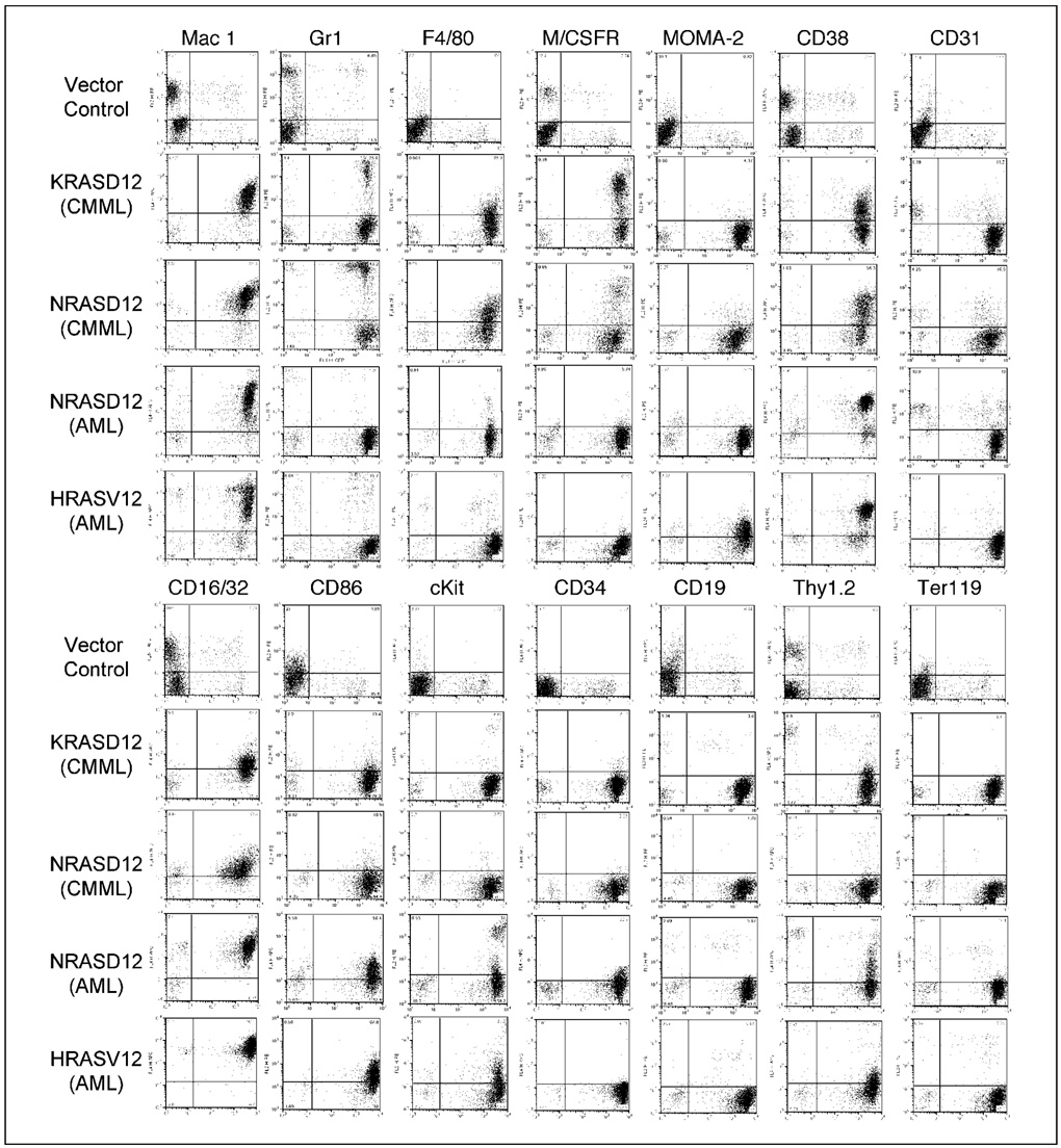
RAS proteins are small GTPases that play a central role in transducing signals that regulate cell proliferation, survival, and differentiation. The RAS proteins interact with a common set of activators and effectors; however, they associate with different microdomains of the plasma membrane as well as other endomembranes and are capable of generating distinct signal outputs. Mutations that result in constitutive activation of RAS proteins are associated with approximately 30% of all human cancers; however, different RAS oncogenes are preferentially associated with different types of human cancer. In myeloid malignancies, NRAS mutations are more frequent than KRAS mutations, whereas HRAS mutations are rare. The mechanism underlying the different frequencies of RAS isoforms mutated in myeloid leukemia is not known. In this study, we compared the leukemogenic potential of activated NRAS, KRAS, and HRAS in the same bone marrow transduction/transplantation model system. We found that all three RAS oncogenes have the ability to induce myeloid leukemias, yet have distinct leukemogenic strengths and phenotypes. The models established here provide a system for further studying the molecular mechanisms in the pathogenesis of myeloid malignancies and for testing targeted therapies.


Figures

Figure 1
Figure 1. Retroviral vectors and RAS expression
A, diagrammatic representation of retroviral vectors used in this study. Bicistronic vectors were used that could coexpress RAS and GFP. Vector control expresses GFP alone. Positions of restriction enzyme sites as well as probe used for Southern blot are indicated. B, Western blot analysis of lysates prepared from NIH3T3 cells infected with NRAS, KRAS, and HRAS retroviruses, as well as the vector control (lanes 1, 2, 3, and 4, respectively) using anti-panRAS and Myc-tag antibodies. Detection of dynamin using an anti-dynamin antibody was used as a loading control.
Figure 2
Figure 2. Cumulative survival of oncogenic NRAS, KRAS, and HRAS mice
Cumulative survival curves of mice transplanted with oncogenic NRAS, KRAS, and HRAS or vector control (MIG) transduced bone marrow cells were generated by Kaplan-Meier survival analysis. Donor bone marrow cells were transduced under closely matched titers of NRAS, KRAS, and HRAS12 retrovirus: ~4 × 10⁵ TU/mL for all RAS and 5 × 10⁶ TU/mL for KRASD12 high titer (HT) and vector control.
Figure 3
Figure 3. Immunophenotyping of leukemic cells from diseased oncogenic NRAS, KRAS, and HRAS mice
Bone marrow or peripheral blood cells were isolated from diseased mice, stained with antibodies to various cell surface markers as indicated, and subjected to flow cytometry analysis. GFP expression is along the X axis, and Y axis shows expression of the cell surface marker specified over each column.
Figure 4
Figure 4. Invasiveness of NRAS- and HRAS-induced AML-like disease in mice
Paraffin sections of lung tissue from vector control and diseased NRAS and HRAS mice were stained with H&E. Images are magnified as indicated.
Figure 5
Figure 5. Proviral integration in leukemic cells from oncogenic NRAS, KRAS, and HRAS mice
A, genomic DNA isolated from leukemic cells of diseased RAS mice (NRAS CMML: lanes 2–3, NRAS AML: lanes 4–6, HRAS: lanes 7–9, KRAS: lanes 10–13) was digested with BglII to analyze proviral integration. A 0.7 kb 32P-labeled GFP fragment was used as the probe. Lane 1 is a control to establish levels of a single copy of the provirus. The blot was stripped and reprobed with a 1.4-kb 32P-labeled irf4 probe as loading control. B, genomic DNA was digested with XbaI (which cuts within the LTRs) to show the intact provirus. Top, MSCV-BCR/ABL-IRES-GFP single-copy proviral control (larger in size than RAS provirus). Bottom, loading control.
Figure 6
Figure 6. Expression levels of oncogenic RAS proteins and activation of downstream pathways
A, myeloid precursor cell line 32Dcl3 cells transduced with vector control (MIG), NRASD12 (N), KRASD12 (K), HRASV12 (H), and BCR/ABL (p210) were starved for 12 h, lysed, and separated on a 6% to 18% polyacrylamide gel. After transfer, the membrane was probed for proteins downstream of RAS, as indicated. B, membrane was also probed for levels of RAS expression, with dynamin as loading control. Ratios of oncogenic RAS to dynamin are indicated. C, liver lysates from leukemic NRAS, KRAS, and HRAS mice were separated on a 6% to 18% gradient gel and probed with anti-RAS antibody.

References

    1. Campbell SL, Khosravi-Far R, Rossman KL, Clark GJ, Der CJ. Increasing complexity of Ras signaling. Oncogene. 1998;17:1395–1413. - PubMed
    1. Khosravi-Far R, Campbell S, Rossman KL, Der CJ. Increasing complexity of Ras signal transduction: involvement of Rho family proteins. Adv Cancer Res. 1998;72:57–107. - PubMed
    1. Hancock JF. Ras proteins: different signals from different locations. Nat Rev Mol Cell Biol. 2003;4:373–384. - PubMed
    1. Rocks O, Peyker A, Bastiaens PI. Spatio-temporal segregation of Ras signals: one ship, three anchors, many harbors. Curr Opin Cell Biol. 2006;18:351–357. - PubMed
    1. Voice JK, Klemke RL, Le A, Jackson JH. Four human ras homologs differ in their abilities to activate Raf-1, induce transformation, and stimulate cell motility. J Biol Chem. 1999;274:17164–17170. - PubMed

Publication types