. 2007 Aug 1;6(15):1878-85.
doi: 10.4161/cc.6.15.4482. Epub 2007 May 21.

Role of survivin phosphorylation by aurora B in mitosis

Affiliations

Role of survivin phosphorylation by aurora B in mitosis

Marlène Delacour-Larose et al. Cell Cycle. .

Abstract

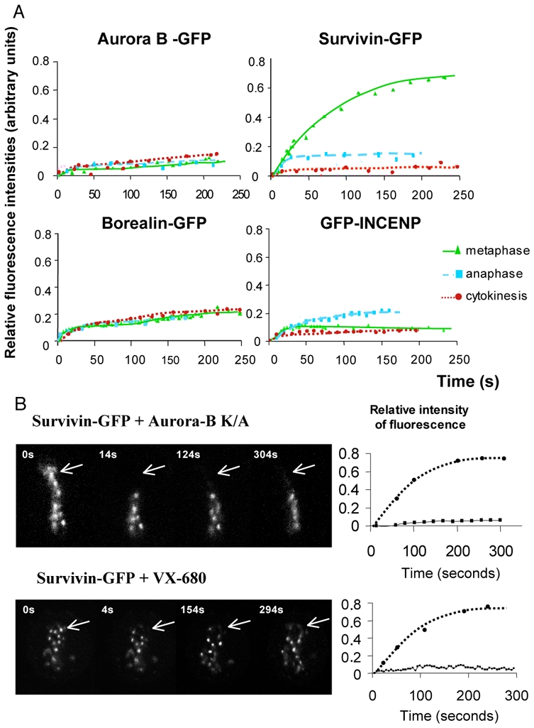
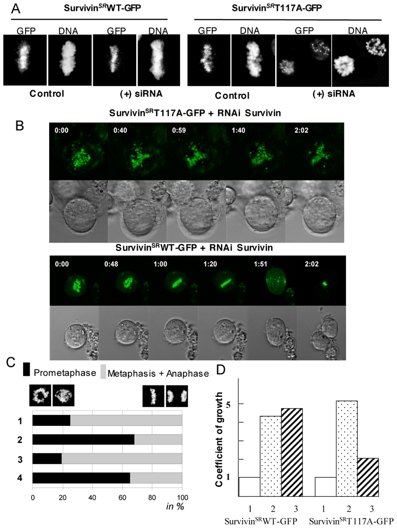
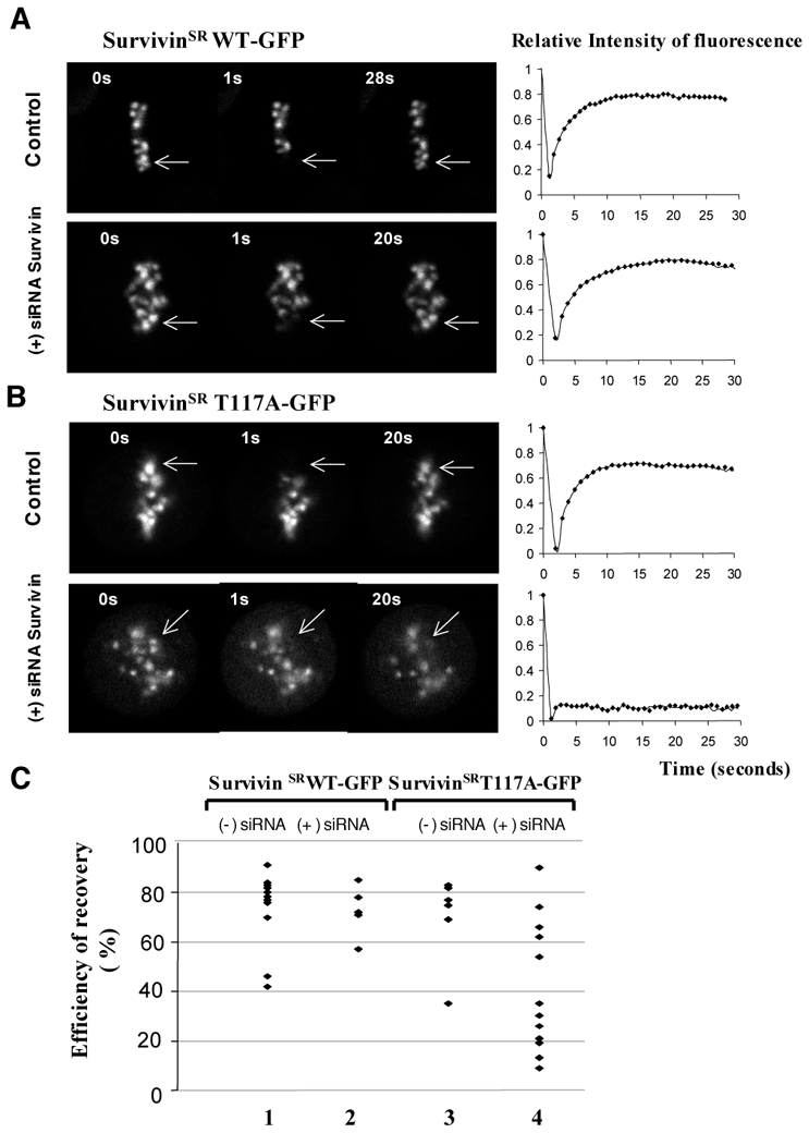
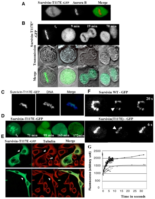
The chromosomal protein passenger complex, a key mitotic regulator, consists of at least four proteins, INCENP, Aurora B, Survivin and Borealin. Survivin, in contrast to the other members of the chromosomal protein passenger complex (CPC), is mobile at metaphase. This protein is also phosphorylated by Aurora B at Threonine 117. In this work we have studied the role of the phosphorylation of Survivin in mitosis by using non phosphorylable T117A and phosphomimic T117E silent resistant Survivin mutants, inducible cell lines expressing these mutants and a combination of siRNA, time-lapse microscopy and FRAP analysis. Time lapse microscopy and FRAP analysis show that Survivin T117A mutant is very stably associated with centromeres and its expression induces a prometaphasic arrest in endogenous survivin depleted cells. In addition, Survivin T117A was unable to rescue the phenotypes of the endogenous survivin depleted cells. Expressed in these cells, the phosphomimic Survivin T117E mutant exhibits a very weak interaction with the centromeres and behaves as a dominant negative mutant inducing severe mitotic defects. Our data suggest that the Aurora B generated phosphorylation/dephosphorylation cycle of Survivin is required for proper proceeding of mitosis.


Figures

Figure 1
Figure 1. FRAP analysis of the passenger proteins mobility during mitosis
(A) HeLa cells were stably transfected with either Aurora B-GFP or Survivin–GFP or transitory transfected with either Borealin-GFP or GFP-INCENP. Either few centromeres at metaphase or a restricted area of the midzone at anaphase and cytokinesis were photobleached. The recovery of fluorescence was measured on the sequence of images acquired at the indicated intervals postbleaching. Note that Survivin, in contrast to the remaining passenger proteins, is highly mobile at metaphase. (B) The mobility of Survivin is dependent on an active Aurora kinase. HeLa cells expressing stably Survivin-GFP were either transfected with an inactive Aurora B kinase (Aurora B K109A) or treated overnight with the specific Aurora B kinase inhibitor, VX-680 at a 300 nM concentration. Few centromeres, indicated by an arrow, were photobleached. The kinetics of recovery of fluorescence are shown in the right part of the figure.
Figure 2
Figure 2. Ectopically expressed Survivin T117A mutant did not restore wild type Survivin function
A) HeLa T-rex cells containing stably integrated tetracyclin inducible expression vectors for either silent resistant wild type Survivin (SurvivinSRWT-GFP, left panel) or silent resistant mutant Survivin (SurvivinSRT117A-GFP, right panel) were treated with Survivin siRNA for 48 hours and the expression of the exogenous Survivin proteins was induced by addition of tetracycline. 16 hours after the tetracycline addition, cells were analysed by microscopy. DNA was stained with Hoechst 33342. Note that the siRNA treated cells expressing SurvivingSRT117A-GFP were arrested at prometaphase. B) GFP-fluorescence and transmission time-lapses imaging of endogenous Survivin depleted cells expressing either SurvivinSRT117A-GFP (top) or SurvivinSRWT-GFP (bottom). Note that the cells expressing SurvivinSRWT-GFP proceed normally in mitosis while the SurvivingSRT117A-GFP are arrested at prometaphase. C) Quantifications of the data. 1, control HeLa T-rex cells; 2, Survivin siRNA treated HeLa T-rex cells; 3, Survivin siRNA treated HeLa T-rex cells expressing SurvivinSRWT-GFP; 4, Survivin siRNA treated HeLa T-rex cells expressing SurvivinSRT117A-GFP. The data represent the average of three independent experiments 100 mitotic cells were scored in each experiment. The percentage of prometaphase (black) and this of the sum of metaphase and anaphase (grey) cells are shown. D) The expression of SurvivinSRT117A-GFP in endogenous Survivin-depleted cells affects cell growth. The same number of cells treated as described in (A) was seeded and the growth rate was estimated 84 hours post seeding. Punctuated rectangles (2) and striated rectangles (3) represent the coefficient of growth (the fold increase of the number of initially seeded cells) for control and Survivin siRNA treated cells, expressing either SurvivinSRWT-GFP (left) or SurvivinSRT117A-GFP (right), respectively. The initial amount of cells was set as one and presented as white rectangles (1).
Figure 3
Figure 3. The substitution of threonine 117 for an alanine residue results in a strong decrease of the mobility of the mutant SurvivinSRT117A-GFP at metaphase
The expression of either SurvivinSRWT-GFP (A) or SurvivinSRT117A-GFP (B) was induced by addition of tetracyclin in either control or Survivin siRNA treated HeLa T-rex cells. The efficiency of expression of both mutatnts was very similar as judged by Western Blotting (data not shown). Few centromeres (indicated by an arrow) of the mitotic cells were photobleached and the recovery of fluorescence was measured at different times postbleaching. The quantification of the FRAP data is shown at the right of each panel. To note is that the kinetics of fluorescence recovery for SurvivingSRT117A-GFP in the siRNA treated cells was decreased to different extent (see panel C) and an extreme example of a quasi-total lost of mobility of SurvivinSRT117A-GFP is shown. C) Summary of the variation of the efficiency of fluorescence recovery (the saturation level of fluorescence recovery measured at 15 seconds postbleaching) in the different FRAP experiments for SurvivinSRWT-GFP and SurvivinSRT117A-GFP in control and Survivin siRNA treated cells. 1 and 2, the efficiency of SurvivinSRWT-GFP fluorescence recovery in control and siRNA treated cells, respectively; 3 and 4, same as 1 and 2, but for the efficiency of SurvivinSRT117A-GFP fluorescence recovery. Each diamond accounts for one cell. Note that SurvivinSRT117A-GFP exhibits very low mobility in a large number of individual Survivin siRNA treated cells.
Figure 4
Figure 4. Localisation and mobility of SurvivinSRT117E-GFP mutant
HeLa T-rex cells were transiently transfected with SurvivinSRT117E-GFP expression vector and the expression of SurvivinSRT117E-GFP was induced by tetracyclin. A) SurvivinSRT117E-GFP loose its centromeric localization in paraformaldehyde fixed cells. HeLa T-rex cells expressing SurvivinSRT117E-GFP were fixed with parafolmaldehyde and the localization of SurvivinSRT117E-GFP was visualized by the fluorescence of its GFP. Aurora B was detected by immunofluorescence by using specific antibodies. DNA was stained with Hoechst 33242. Typical example of a metaphasic cell is shown. B) GFP-fluorescence and transmission time lapse imaging of SurvivinSRT117E-GFP. The merge of both signals is also presented. Note that SurvivinSRT117E-GFP is localized at the chromosomes in metaphase, but is not transferred to the central spindle and the midbody as mitosis proceeds. C) SurvivinSRT117E-GFP is associated with mitotic chromosomes. SurvivinSRT117E-GFP was visualized by the GFP-fluorescence, while DNA was stained with Hoechst 33242. Hoechst 33242 was added for 10 min in the culture medium. Following a quick wash with culture medium, the cells were immediately imaged at 488 nm (GFP) and 720 nm (Hoechst). The merge of both signals is also shown. D) Expression of SurvivinSRT117E-GFP results in a complex cell phenotype. A Hela T-rex cell expressing SurvivinSRT117E-GFP was continuously imaged. This time-lapse reveals the formation of a polyploidy cell within 3 hours. E) Immunofluorescence microscopy of fixed cells expressing SurvivinSRT117E-GFP. Cells were fixed and submitted to an IF. Green, SurvivinSRT117E-GFP; red, tubulin, detected with a specific anti-tubulin antibody. Single arrowheads point abnormal connection between cells, whereas double arrowhead point a normal mid-body in non transfected cells. F) SurvivinSRT117E-GFP is highly mobile at metaphase. HeLa T-rex cells were transiently transfected with either SurvivinSRWT-GFP or SurvivinSRT117E-GFP expression vectors and the protein expression was induced by tetracyclin. Few centromeres were bleached and fluorescence recovery was imaged. G) FRAP quantifications. FRAP were performed as in F except that a rapid scanning of fluorescence recovery was performed by imaging a Region Of Interest (ROI) including the bleached region. Times between two scanning was 60 ms. The squares (dotted line) represented the chimera SurvivinSR-GFP and the diamonds (continuous line) the SurvivinSRT117E-GFP.

References

    1. Adams RR, Carmena M, Earnshaw WC. Chromosomal passengers and the (aurora) ABCs of mitosis. Trends Cell Biol. 2001;11:49–54. - PubMed
    1. Honda R, Korner R, Nigg EA. Exploring the functional interactions between Aurora B, INCENP, and survivin in mitosis. Mol Biol Cell. 2003;14:3325–3341. - PMC - PubMed
    1. Andrews PD, Ovechkina Y, Morrice N, Wagenbach M, Duncan K, Wordeman L, Swedlow JR. Aurora B regulates MCAK at the mitotic centromere. Dev Cell. 2004;6:253–268. - PubMed
    1. Hirota T, Lipp JJ, Toh BH, Peters JM. Histone H3 serine 10 phosphorylation by Aurora B causes HP1 dissociation from heterochromatin. Nature. 2005;438:1176–1180. - PubMed
    1. Nowak SJ, Corces VG. Phosphorylation of histone H3: a balancing act between chromosome condensation and transcriptional activation. Trends Genet. 2004;20:214–220. - PubMed

Publication types