. 2007 Aug;9(8):858-67.
doi: 10.1038/ncb0807-858.

Functional atlas of the integrin adhesome

Affiliations

Functional atlas of the integrin adhesome

Ronen Zaidel-Bar et al. Nat Cell Biol. 2007 Aug.

Abstract

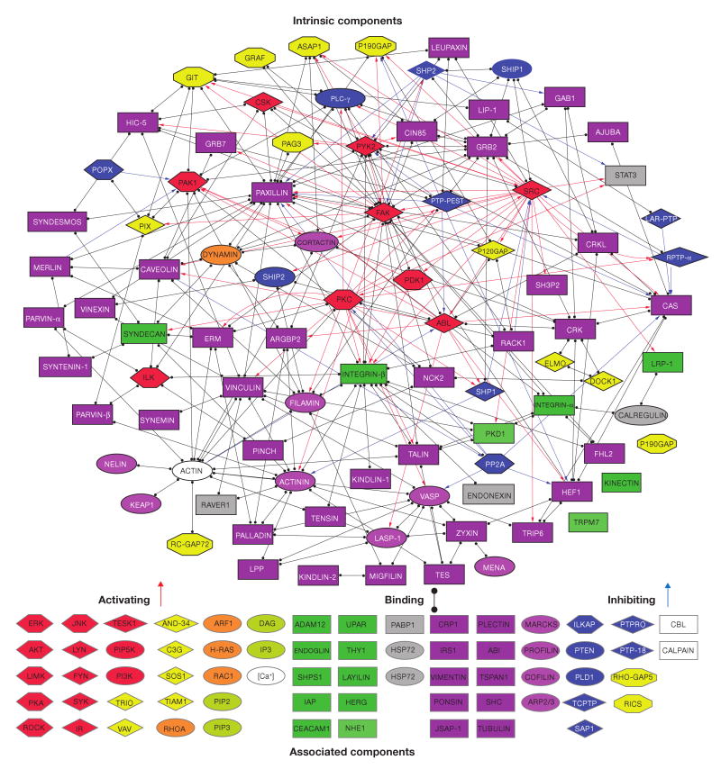
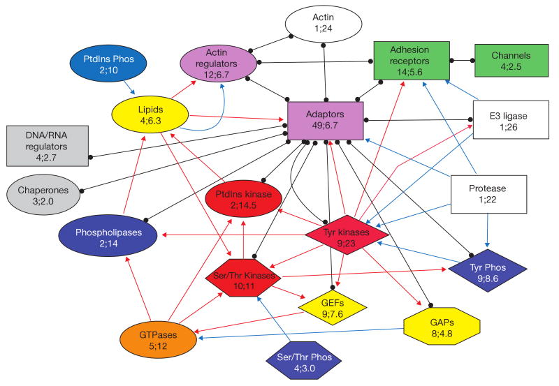
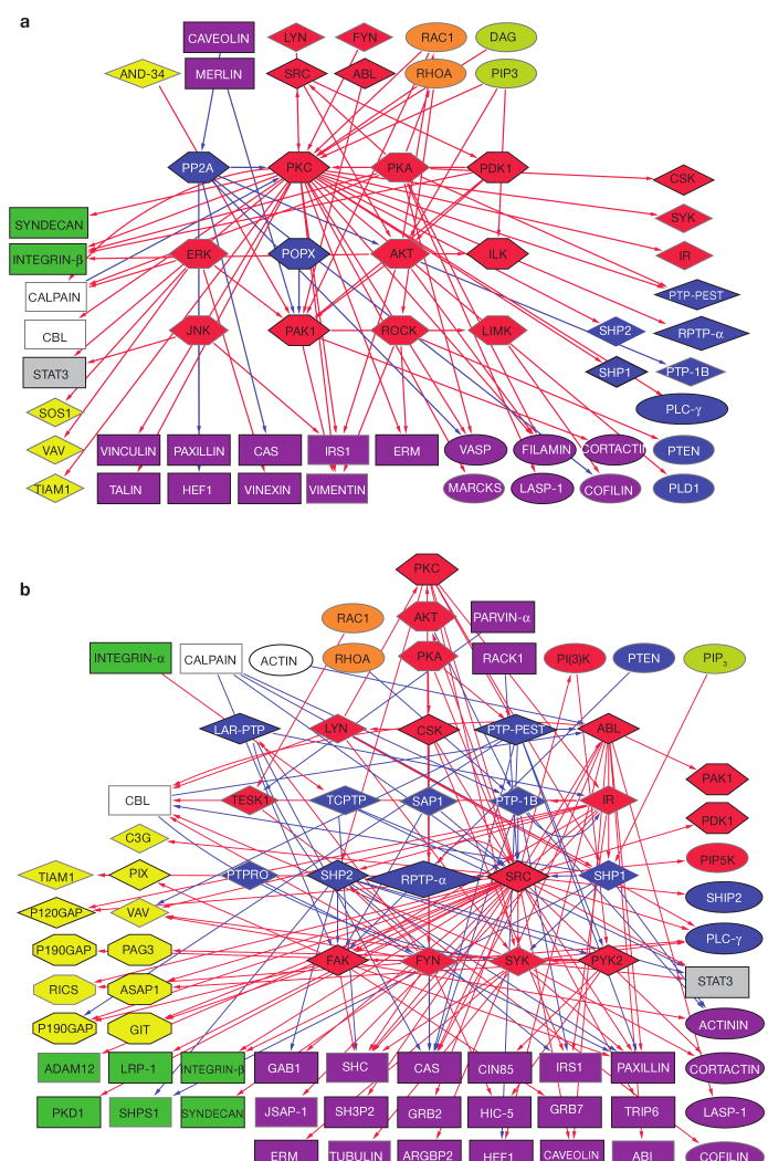
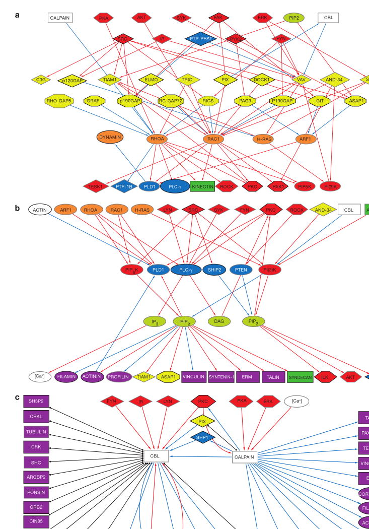
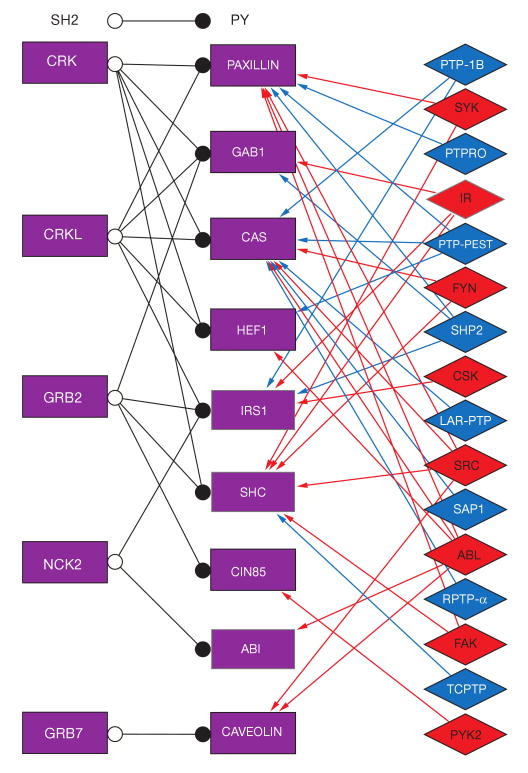
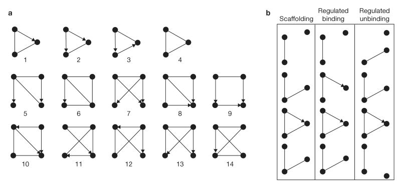
A detailed depiction of the 'integrin adhesome', consisting of a complex network of 156 components linked together and modified by 690 interactions is presented. Different views of the network reveal several functional 'subnets' that are involved in switching on or off many of the molecular interactions within the network, consequently affecting cell adhesion, migration and cytoskeletal organization. Examination of the adhesome network motifs reveals a relatively small number of key motifs, dominated by three-component complexes in which a scaffolding molecule recruits both a signalling molecule and its downstream target. We discuss the role of the different network modules in regulating the structural and signalling functions of cell-matrix adhesions.


Figures

Figure 1
Figure 1
Interactions between all intrinsic components of the adhesome and a grouped list of the associated components. Black lines with full circles at their ends denote non-directional binding interactions, blue arrows represent directional inhibition (for example, dephosphorylation, G-protein inactivation or proteolysis) and red arrows represent directional activation (for example, phosphorylation or G-protein activation) interactions. The nodes are shape- and colour-coded according to the function of the proteins, as detailed in Fig. 2. Intrinsic components are surrounded by a black frame and associated components by a grey frame. For details see supplementary information, S2.
Figure 2
Figure 2
Interactions between functional families of adhesome components. Each protein in the adhesome was categorized into one of 20 groups according to its known biological activity. The families are shown in unique combinations of colour and shape, indicating the number of family members followed by the average number of their interactions. In addition, the dominating interactions between families (red arrows, activating interactions; blue arrows, inhibiting interactions; black lines, binding interactions) are shown. For details see supplementary information, S2.
Figure 3
Figure 3
Actin–integrin subnet interconnecting the membrane receptors (mainly integrin) with actin. Only binding interactions are shown. This subnet consists of actin, actin modulators (11 molecules), adaptor proteins (46 molecules) and transmembrane molecules (mainly integrin receptors). For details see supplementary information, S2.
Figure 4
Figure 4
Phosphorylation subnets mapping serine/threonine kinases and phosphatases (a) and tyrosine kinases and phosphatases (b). The figure depicts the corresponding kinases and phosphatases (in the centre), their diverse substrates (bottom and sides) and their regulators (top). The schematic shows hubs of activities (for example, PKC, Src) and a wide variety of substrates. For details see supplementary information, S2.
Figure 5
Figure 5
GTPase, lipid and proteolytic subnets. (a) The G-proteins associated with the adhesome (mainly RhoA and Rac1) are shown above their main substrates and below their direct positive (GEFs) and negative (GAPs) regulators. Regulators of the GEFs and GAPs are mainly kinases (top). (b) Signalling lipids are shown with their diverse substrates, the regulatory kinases and phosphatases and their regulators (mainly kinases and G-proteins). (c) The two proteolytic systems associated with the adhesome (Cbl and calpain) affect multiple substrates, and are affected mainly by tyrosine kinases (Cbl) or serine/threonine kinases (calpain). For details see supplementary information, S2.
Figure 6
Figure 6
Phosphorylation switches regulating specific phosphotyrosine–SH2 domain interactions. The adaptor proteins on the left contain SH2 domains, which can bind to phosphotyrosine residues present on the adaptor proteins in the centre. The interaction between the pairs of adaptor proteins is regulated by the phosphorylation state of the tyrosine residues, which depends on the activity of the tyrosine kinases or phosphatases shown on the right. For details see supplementary information, S2.
Figure 7
Figure 7
Network motifs of the adhesome. (a) A schematic representation of the most significant three-node and four-node network motifs found in the adhesome. The statistical significance of each of these is listed in Table 1. (b) Three possibilities for the dynamic assembly or disassembly of a protein complex, based on interactions such as those depicted in motif 1. This motif acts as a signalling scaffold when binding of the substrate to the third protein precedes the enzymatic reaction: for example, the binding of PAK1 to PIX is a pre-requisite for its dephosphorylation by POPX, which is also bound to PIX. In other cases, the binding of the substrate to the third protein is positively or negatively regulated by the enzymatic reaction. An example of positive regulation would be the binding of HEF1 to paxillin only after it is phosphorylated by PYK2, which is also bound to paxillin. An example for negative regulation would be when SHP2 dephosphorylates IRS1 and breaks its binding to GRB2.

Similar articles

Cited by

References

    1. Burridge K, Fath K, Kelly T, Nuckolls G, Turner C. Focal adhesions: transmembrane junctions between the extracellular matrix and the cytoskeleton. Annu Rev Cell Biol. 1988;4:487–525. - PubMed
    1. Geiger B, Bershadsky A, Pankov R, Yamada KM. Transmembrane crosstalk between the extracellular matrix — cytoskeleton crosstalk. Nature Rev Mol Cell Biol. 2001;2:793–805. - PubMed
    1. Critchley DR, et al. Integrin-mediated cell adhesion: the cytoskeletal connection. Biochem Soc Symp. 1999;65:79–99. - PubMed
    1. Geiger B, Bershadsky A. Exploring the neighborhood: adhesion-coupled cell mechanosensors. Cell. 2002;110:139–142. - PubMed
    1. Bershadsky A, Kozlov M, Geiger B. Adhesion-mediated mechanosensitivity: a time to experiment, and a time to theorize. Curr Opin Cell Biol. 2006;18:472–481. - PubMed

Publication types