

Comment
doi: 10.1172/JCI32966.
Proteinuria: is it all in the foot?
Affiliations
- PMID: 17671644
- PMCID: PMC1934599
- DOI: 10.1172/JCI32966
Item in Clipboard
Comment
Proteinuria: is it all in the foot?
J Clin Invest.
2007 Aug.
Abstract
Despite significant advances in our understanding of the molecular structure and composition of the glomerular filtration barrier, the mechanisms underlying the presence of excess protein in the urine (proteinuria) in acquired human kidney diseases remain elusive. In a study appearing in this issue of the JCI, Sever and associates use a combination of biochemical, genetic, and in vivo approaches in mice to demonstrate a pivotal role of cathepsin L and its substrate the GTPase dynamin, in the induction of proteinuria and associated foot process effacement in glomerular podocytes (see the related article beginning on page 2095).
Figures
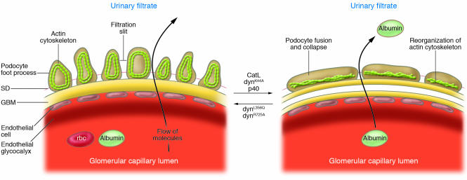
Blood enters the glomerular capillaries and is filtered across the endothelium and the glomerular basement membrane and through the filtration slits between podocyte foot processes to produce the primary urine filtrate. In healthy glomeruli, this barrier restricts the passage of macromolecules. In this issue of the JCI, Sever et al. (10) show that CatL, the expression of which is increased in human proteinuric diseases and in an LPS-induced mouse model of proteinuria, causes proteinuria and foot process effacement through cleavage of the GTPase dynamin, an actin-binding protein. The same effects are induced by gene delivery into mice of dynK44A — a mutant form of dynamin that does not bind GTP — or of the CatL-cleaved product of dynamin (p40). Conversely, gene delivery into proteinuric mice of dynL356Q and dynR725A, two CatL-resistant dynamin mutants, reverses proteinuria and foot process effacement. Figure modified from ref. .
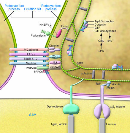
This schematic shows the connections between the cortical actin cytoskeleton and components of the basolateral portion of a podocyte. The membrane domains are marked: apical cell membrane (green); the SD domain (blue); and the “sole” of the foot process (pink). In this issue of the JCI, Sever et al. (10) show that LPS injection into mice induces CatL expression in the cytoplasm, which cleaves the active form of GTPase dynamin (represented here in its GTP-bound homotetrameric form). Dynamin binds actin-regulatory proteins including cortactin and the Arp2/3 complex, which catalyze actin filament assembly. Cleavage of dynamin generates a 40-kDa dynamin fragment (p40), which interferes with the normal function of dynamin and induces cytoskeleton reorganization, foot process effacement, and proteinuria as shown in Figure 1. Figure adapted from ref. .
Comment on
-
Proteolytic processing of dynamin by cytoplasmic cathepsin L is a mechanism for proteinuric kidney disease.J Clin Invest. 2007 Aug;117(8):2095-104. doi: 10.1172/JCI32022. J Clin Invest. 2007. PMID: 17671649 Free PMC article.
References
-
- Kestila M., et al. Positionally cloned gene for a novel glomerular protein — nephrin — is mutated in congenital nephrotic syndrome. Mol. Cell. 1998;1:575–582. - PubMed
-
- Tryggvason K., Patraaka J., Wartiovaara J. Hereditary proteinuria syndromes and mechanisms of proteinuria. N. Engl. J. Med. 2006;354:1387–1401. - PubMed
-
- Boute N., et al. NPHS2, encoding the glomerular protein podocin, is mutated in autosomal recessive steroid-resistant nephrotic syndrome. Nat. Genet. 2000;24:349–354. - PubMed
-
- Huber T.B., et al. Interaction with podocin facilitates nephrin signaling. J. Biol. Chem. 2001;276:41543–41546. - PubMed
