. 2007 Aug;117(8):2095-104.
doi: 10.1172/JCI32022.

Proteolytic processing of dynamin by cytoplasmic cathepsin L is a mechanism for proteinuric kidney disease

Affiliations

Proteolytic processing of dynamin by cytoplasmic cathepsin L is a mechanism for proteinuric kidney disease

Sanja Sever et al. J Clin Invest. 2007 Aug.

Abstract

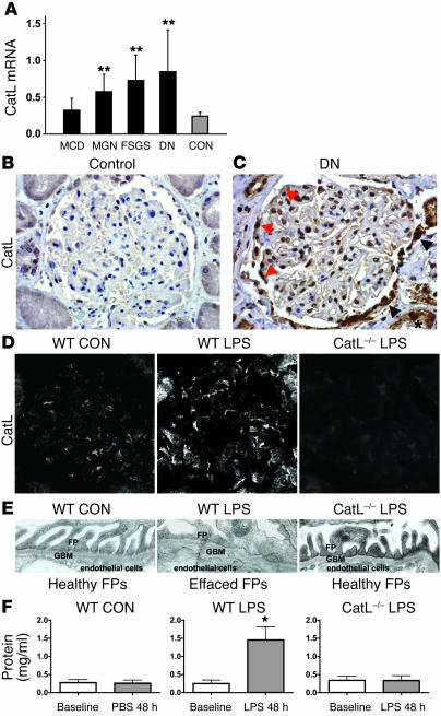
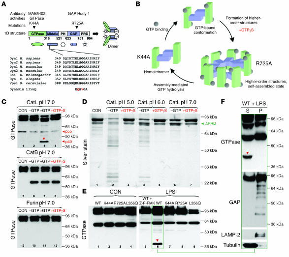
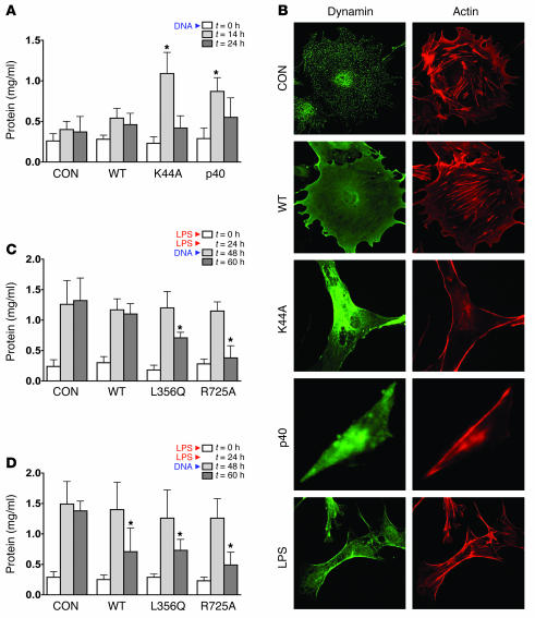
Kidney podocytes and their foot processes maintain the ultrafiltration barrier and prevent urinary protein loss (proteinuria). Here we show that the GTPase dynamin is essential for podocyte function. During proteinuric kidney disease, induction of cytoplasmic cathepsin L leads to cleavage of dynamin at an evolutionary conserved site, resulting in reorganization of the podocyte actin cytoskeleton and proteinuria. Dynamin mutants that lack the cathepsin L site, or render the cathepsin L site inaccessible through dynamin self-assembly, are resistant to cathepsin L cleavage. When delivered into mice, these mutants restored podocyte function and resolve proteinuria. Our study identifies dynamin as a critical regulator of renal permselectivity that is specifically targeted by proteolysis under pathological conditions.


Figures

Figure 1
Figure 1. CatL is essential for proteinuric kidney diseases.
(A) Quantitative rt-PCR of microdissected glomeruli from human biopsies of patients with acquired proteinuric diseases: minimal change disease (MCD; n = 7), membranous glomerulonephritis (MGN; n = 9), focal segmental glomerulosclerosis (FSGS; n = 7), and diabetic nephropathy (DN; n = 10). **P < 0.01. CON, control (n = 8). (B) CatL labeling of normal human kidney. (C) CatL labeling of human kidney with diabetic nephropathy, mildly reduced renal function, and nephrotic range proteinuria. (D) Immunocytochemistry of mouse glomeruli using monoclonal anti-CatL antibody. WT mice received either PBS (WT CON) or LPS (WT LPS). LPS was also injected into CatL–/– mice (CatL–/– LPS). Original magnification, ×400 (C and D). (E) Electron micrographs of FPs. (F) Urinary protein levels determined using the standard Bradford protein assay. Urine was collected immediately before (Baseline) and 48 hours after addition of LPS. Each bar represents at least 8 animals.
Figure 2
Figure 2. Induction of cytoplasmic CatL protein and activity by LPS.
(A) Cultured podocytes were stained using anti–LAMP-2 antibodies and the BIOMOL CV-CatL/B activity detection kit. Control cells, cells treated with 50 μg/ml of LPS for 24 hours, and cells treated with LPS and 1 μM of the selective CatL inhibitor Z-FF-FMK are shown. (B) Labeling of cultured podocytes using anti-CatL antibody, untreated or after treatment with 50 μg/ml of LPS for 24 hours. (C) Labeling of cultured podocytes using anti-CatB antibody, untreated or after treatment with 50 μg/ml of LPS for 24 hours. (D) Schematic of CatL mRNA and resulting proteins. (E) Subcellular fractionation of podocytes in isotonic sucrose prior to and 24 hours after LPS treatment. Total proteins from the soluble (S) and the particulate (P) fractions were analyzed by Western blotting.
Figure 3
Figure 3. Altered dynamin staining in glomerulus after LPS treatment is CatL dependent.
(A) Immunogold analysis of dynamin in podocyte FPs of rat glomerulus. Low-power view (left) depicts dynamin within the center of podocyte FPs (asterisk) as well as in electron-dense areas rich of cortical actin (a) (original magnification, ×30,000). High-power view shows gold particles, which are associated with the cytoplasmic side of vesicles (v) and with electron-dense actin areas (a) (original magnification, ×45,000). E, endosome; GBM, glomerular basement membrane; Ly, lysosome; SD, slit diaphragm; US, urinary space. (B) Immunocytochemistry of dynamin in glomeruli of WT mice before and after injection of LPS and of CatL–/– mice after injection of LPS. (C) Immunocytochemistry of samples from CatL–/– mice for glomerular dynamin and CatL. CatL–/– mice had been reconstituted with either Pre-Pro-CatL (left) or with short CatL (right) using transient gene transfer. Twenty-four hours after gene transfer, glomeruli were stained using anti-CatL and anti-dynamin hudy 1 antibody. Original magnification, ×400 (B and C). (D) Immunoblot of endogenous dynamin in cultured podocytes in response to LPS. Recombinant dynamin (REC), extracts of WT and CatL–/– podocytes, were analyzed. (E) Quantitation of dynamin in cell extracts shown in D. Dynamin signal was adjusted to actin levels.
Figure 4
Figure 4. Effects of the nucleotide-bound and assembly state of dynamin on CatL cleavage in vitro and in vivo.
(A) Domain structure of dynamin, corresponding antibodies, and amino acid sequence of predicted CatL cleavage sites. Note that the ELSGGA sequence is a highly conserved motif throughout the species. PH, pleckstrin homology domain; PRD, proline-arginine rich domain; Shi, dynamin homolog in Drosophila; Vsp1, dynamin homolog in yeast. (B) Schematic depiction of dynamin GTPase cycle. In its basal state, dynamin is a homotetramer. Self-assembly into higher-order structures such as rings or spirals can be promoted by GTPϒS and activates assembly-mediated GTP hydrolysis, which in turn drives disassembly. Dynamin’s middle domain is located inside the spiral. (C) Recombinant dyn1 (20 pmol) (CON) was mixed with CatL (1 pmol) (top panel), CatB (middle panel), or Furin (bottom panel). The reactions were performed at pH 7.0 under nonassembly conditions (200 mM NaCl). Where indicated, 200 μM GTP or 1 mM GTPϒS was present. Proteolytic products were detected by monoclonal anti-dynamin antibody against the GTPase domain. (D) Silver staining of recombinant dyn1 incubated with CatL at different pHs in the presence or absence of GTPϒS. (E) Western blot analysis using GTPase antibody of podocyte extracts infected with various dynamin mutants before or after addition of 100 μg/ml LPS for 20 hours. Note the appearance of a 40-kDa fragment (arrow). When indicated, podocytes were treated with 1 μM of the selective CatL inhibitor Z-FF-FMK for the duration of the LPS treatment. (F) Western blot analysis of subcellular fractionation of podocytes expressing dynWT for 24 hours after LPS treatment. Extracts were blotted using antibodies against the GTPase domain (N-terminal), GAP domain (C-terminal), LAMP-2, and tubulin.
Figure 5
Figure 5. LPS-induced dynamin cleavage in mouse kidneys.
(AC) Double immunofluorescence of dyn1 (MAB-5402 antibody; red) and the podocyte marker synaptopodin (green). Mice were injected with podocin-driven expression vector lacking the dynamin gene (A), CMV-dyn1 expression vector (B), and POD-dyn1 expression vector (C). Western blot analysis of total kidney and liver extracts isolated from animals injected with different cDNAs as indicated in the figure. (D) Immunocytochemistry of glomeruli from mice that were injected with LPS. Twenty-four hours after LPS injections, animals received cDNA expressing dynWT, dynL356Q, and dynR725A and were sacrificed 14 hours later. Glomeruli were stained using monoclonal anti-dynamin hudy 1 antibody. Original magnification, ×400 (AD). (E) Western blot analysis of kidney extracts after gene delivery of different dyn1 cDNAs using the GTPase and CatL antibody before and after addition of LPS.
Figure 6
Figure 6. The effects of dynamin mutants on proteinuria and podocyte morphology.
(A) Urinary protein levels determined using the standard Bradford protein assay. Urine was collected immediately before (0 h) and 14 and 24 hours after injection of podocin-driven expression vectors containing different dynamin constructs as indicated in the figure. Each bar represents at least 10 animals. (B) Cultured podocytes were infected with adenoviruses expressing the indicated dynamin constructs. Eighteen hours after infection, cells were stained with hudy 1 (green) to visualize dynamin and rhodamine phalloidin (red) to visualize F-actin. (C) Urinary protein levels determined using the standard Bradford protein assay. Mice were injected at 0 and 24 hours with LPS to induce proteinuria. At 48 hours (peak proteinuria), mice received empty vector, CMV-dynWT, CMV-dynL356Q, or CMV-dynR725A. Urinary protein levels were determined 12 hours after injection. Each data point represents at least 10 animals. (D) Urinary protein levels determined using the standard Bradford protein assay. Mice were injected at 0 and 24 hours with LPS to induce proteinuria. At 48 hours (peak proteinuria), mice received empty vector, POD-dynWT, POD-dynL356Q, or POD-dynR725A. Urinary protein levels were determined 12 hours after injection. Each bar represents at least 10 animals.

Comment in

References

    1. Zandi-Nejad K., Eddy A.A., Glassock R.J., Brenner B.M. Why is proteinuria an ominous biomarker of progressive kidney disease? . Kidney Int. Suppl. 2004;66:S76–S89. - PubMed
    1. Tryggvason K., Patrakka J., Wartiovaara J. Hereditary proteinuria syndromes and mechanisms of proteinuria. N. Engl. J. Med. 2006;354:1387–1401. - PubMed
    1. Reiser J., et al. Podocyte migration during nephrotic syndrome requires a coordinated interplay between cathepsin l and alpha3 integrin. J. Biol. Chem. 2004;279:34827–34832. - PubMed
    1. Baricos W.H., O’Connor S.E., Cortez S.L., Wu L.T., Shah S.V. The cysteine proteinase inhibitor, E-64, reduces proteinuria in an experimental model of glomerulonephritis. Biochem. Biophys. Res. Commun. 1988;155:1318–1323. - PubMed
    1. Goulet B., et al. A cathepsin L isoform that is devoid of a signal peptide localizes to the nucleus in S phase and processes the CDP/Cux transcription factor. Mol. Cell. 2004;14:207–219. - PubMed

Publication types

MeSH terms