

Risk factors associated with the loss of cartilage volume on weight-bearing areas in knee osteoarthritis patients assessed by quantitative magnetic resonance imaging: a longitudinal study
- PMID: 17672891
- PMCID: PMC2206376
- DOI: 10.1186/ar2272
Risk factors associated with the loss of cartilage volume on weight-bearing areas in knee osteoarthritis patients assessed by quantitative magnetic resonance imaging: a longitudinal study
Abstract
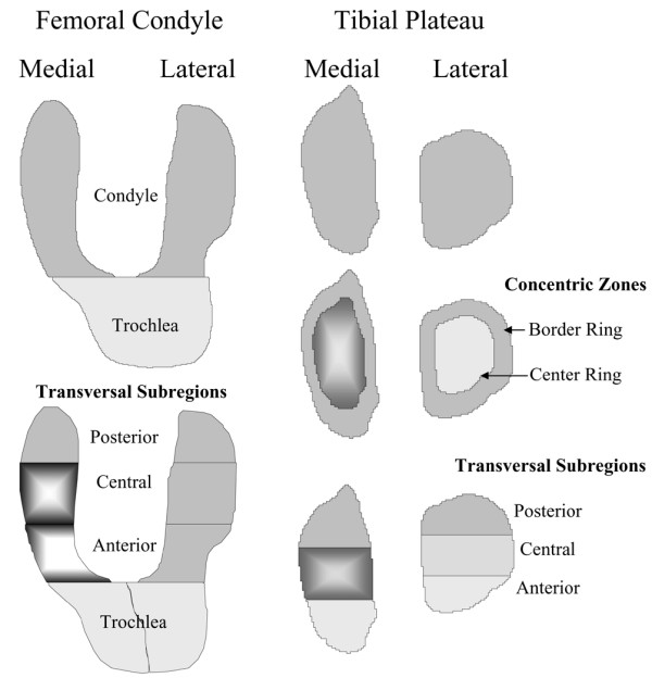
The objective of this study was to identify, on a symptomatic knee osteoarthritis (OA) cohort, the risk factors associated with the progression of the disease. More specifically, we investigated the correlation between knee cartilage volume loss from subregions over the span of 24 months by means of quantitative magnetic resonance imaging (qMRI) with demographic, clinical, radiological, and MRI structural changes. A cohort of 107 patients with knee OA selected from a large trial evaluating the effect of a bisphosphonate underwent x-rays and MRI of the knee at baseline and 24 months. Joint space width (JSW) and joint space narrowing (JSN) and cartilage volume loss over time in subregions of the tibial plateaus and femoral condyles were quantitated. Structural changes in the subchondral bone (hypersignal) and in the menisci (tear and extrusion) were also evaluated. The greatest cartilage volume loss was found in the medial compartment, and risk factors included female gender, JSW, meniscal lesions, and bone changes at baseline. Subregion analysis revealed that the greatest cartilage volume loss at 24 months was found in the central area of the medial tibial plateau (15%; p < 0.0001) and of the medial femoral condyle (12%; p < 0.0001). These findings were associated with the presence at baseline of meniscal extrusion, particularly severe meniscal extrusion, medial and severe meniscal tear, bone hypersignal, high body mass index (BMI), smaller JSW, increases in Western Ontario and McMaster Universities Osteoarthritis Index (WOMAC) pain and patient global scores over time, and greater JSN. Parameters predicting medial central femoral condyle cartilage volume loss at 24 months were lateral meniscal tear, SF-36 and BMI at baseline, and JSN. At the medial central tibial plateau, the parameters were severe meniscal extrusion, severe lateral meniscal tear, and bone hypersignal in the lateral compartment at baseline, and WOMAC pain change. Meniscal damage and bone changes are the features most closely associated with the greatest subregional cartilage volume loss. Interestingly, for the first time, JSN was strongly associated with cartilage loss in the central areas of plateaus and condyles. This study also further confirms the correlation between cartilage volume loss and JSN and symptomatic changes at 24 months.
Figures
References
-
- Martel-Pelletier J, Lajeunesse D, Pelletier JP. Etiopathogenesis of osteoarthritis. In: Koopman WJ, Moreland LW, editor. Arthritis and Allied Conditions: A Textbook of Rheumatology. Baltimore, MD: Lippincott, Williams & Wilkins; 2005. pp. 2199–2226.
-
- Berthiaume MJ, Raynauld JP, Martel-Pelletier J, Labonté F, Beaudoin G, Bloch DA, Choquette D, Haraoui B, Altman RD, Hochberg M, et al. Meniscal tear and extrusion are strongly associated with the progression of knee osteoarthritis as assessed by quantitative magnetic resonance imaging. Ann Rheum Dis. 2005;64:556–563. doi: 10.1136/ard.2004.023796. - DOI - PMC - PubMed
-
- Sowers MF, Hayes C, Jamadar D, Capul D, Lachance L, Jannausch M, Welch G. Magnetic resonance-detected subchondral bone marrow and cartilage defect characteristics associated with pain and X-ray-defined knee osteoarthritis. Osteoarthritis Cartilage. 2003;11:387–393. doi: 10.1016/S1063-4584(03)00080-3. - DOI - PubMed
-
- Amin S, LaValley MP, Guermazi A, Grigoryan M, Hunter DJ, Clancy M, Niu J, Gale DR, Felson DT. The relationship between cartilage loss on magnetic resonance imaging and radiographic progression in men and women with knee osteoarthritis. Arthritis Rheum. 2005;52:3152–3159. doi: 10.1002/art.21296. - DOI - PubMed
-
- Ding C, Garnero P, Cicuttini F, Scott F, Cooley H, Jones G. Knee cartilage defects: association with early radiographic osteoarthritis, decreased cartilage volume, increased joint surface area and type II collagen breakdown. Osteoarthritis Cartilage. 2005;13:198–205. doi: 10.1016/j.joca.2004.11.007. - DOI - PubMed
Publication types
MeSH terms
LinkOut - more resources
Full Text Sources
Other Literature Sources
Medical
