. 2007 Oct 1;313(16):3616-23.
doi: 10.1016/j.yexcr.2007.07.002. Epub 2007 Jul 10.

Activation of ROCK by RhoA is regulated by cell adhesion, shape, and cytoskeletal tension

Affiliations

Activation of ROCK by RhoA is regulated by cell adhesion, shape, and cytoskeletal tension

Kiran Bhadriraju et al. Exp Cell Res. .

Abstract

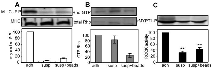
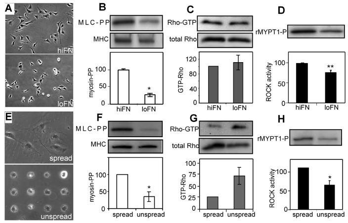
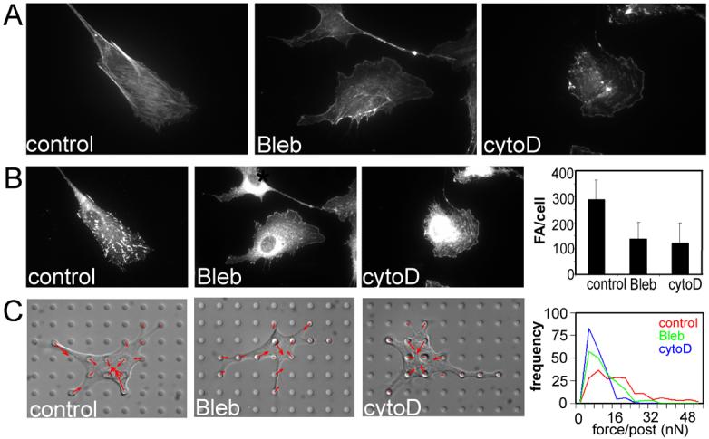
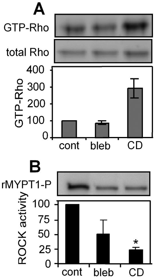
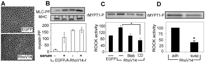
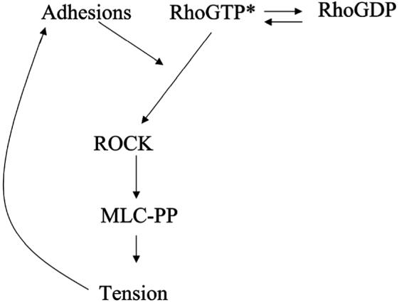
Adhesion to the extracellular matrix regulates numerous changes in the actin cytoskeleton by regulating the activity of the Rho family of small GTPases. Here, we report that adhesion and the associated changes in cell shape and cytoskeletal tension are all required for GTP-bound RhoA to activate its downstream effector, ROCK. Using an in vitro kinase assay for endogenous ROCK, we found that cells in suspension, attached on substrates coated with low density fibronectin, or on spreading-restrictive micropatterned islands all exhibited low ROCK activity and correspondingly low myosin light chain phosphorylation, in the face of high levels of GTP-bound RhoA. In contrast, allowing cells to spread against substrates rescued ROCK and myosin activity. Interestingly, inhibition of tension with cytochalasin D or blebbistatin also inhibited ROCK activity within 20 min. The abrogation of ROCK activity by cell detachment or inhibition of tension could not be rescued by constitutively active RhoA-V14. These results suggest the existence of a feedback loop between cytoskeletal tension, adhesion maturation, and ROCK signaling that likely contributes to numerous mechanochemical processes.


Figures

Fig. 1
Fig. 1
Placing cells in suspension decouples Rho activity from ROCK. A) Western blots (top) and normalized graphs (bottom) of phosphorylated myosin light chain (Ser19/Thr18) and myosin heavy chain in adherent, suspended, and suspended cells incubated with fibronectin-coated beads; n=2. B) Western blots (top) and normalized graphs (bottom) of GTP-Rho and total Rho in adherent, suspended, and suspended cells incubated with fibronectin-coated beads; n=2. C) Western blot (top) and normalized graphs (bottom) of ROCK kinase assay product, phosphoMYPT1 Thr696 adherent, suspended, and suspended cells incubated with fibronectin-coated beads; n=3. Error bars indicate standard errors of mean; ** (p<0.005).
Fig. 2
Fig. 2
ECM density and cell shape regulate Rho-ROCK coupling. A and E: Phase contrast micrograph of cells on high fibronectin or ‘spread’ condition (A,E top, respectively) or low fibronectin (A,E bottom, respectively) substrates. B and F: Western blots (top) and normalized graphs (bottom) of phosphorylated myosin light chain (Ser19/Thr18) and myosin heavy chain in cells on hiFN and loFN (B; n=3) or ‘spread’ or ‘unspread’ condition (F; n=3). C and G: Western blots (top) and normalized graphs (bottom) of GTP-Rho and total Rho in cells on hiFN and loFN (C; n=8) or in ‘spread’ or ‘unspread’ condition (G; n=2 experiments). D and H: Western blot (top) and normalized graphs (bottom) of ROCK kinase assay product, phosphoMYPT1 Thr696 in cells on hiFN and loFN (D; n=6) and ‘spread’ and ‘unspread’ condition (H; n=4). Error bars indicate standard errors of mean; * (p<0.05), ** (p<0.005).
Fig. 3
Fig. 3
The effect of tension inhibiting drugs on stress fibers, focal adhesions and contractility. A) Immunofluorescence micrographs of stress fibers labeled with TRITC-phalloidin in control (left), blebbistatin-treated (25μM, middle), and cytochalasin D-treated (2μg/ml, right) cells. B) Immunofluorescence micrographs of focal adhesions labeled with anti-vinculin antibody in control (left), blebbistatin-treated (middle), and cytochalasin D-treated (right) cells. C) Graphs of quantified number of focal adhesions per cell in drug treated cells compared to controls. D) Differential interference contrast micrographs of control (left), blebbistatin-treated (middle), and cytochalasin D-treated (right) cells on the microfabricated force-measuring device, overlaid with contractile force vectors. The top graph to the right the average number of focal adhesions per cell in control, blebbistatin-treated and cytochalasin D-treated cells. The bottom graphs shows histograms of contractile forces, each histograms representing pooled data from 15 cells under the respective conditions from 3 experiments: red - control cells, green - blebbistatin-treated cells, blue - cytochalasin D treated cells. Error bars indicate standard deviations of measurement of about 50 cells from a representative experiment.
Fig. 4
Fig. 4
Blocking cytoskeletal tension blocks ROCK activity irrespective of GTP-Rho level. A) Western blot (top) and graph of normalized RhoA (bottom) of Rho pull down in control (cont), blebbistatin (bleb) and cytochalasin D (CD)-treated cells; n=2. B) Western blot (top) and graph of normalized ROCK activity (bottom) in control, blebbistatin and cytochalasin D-treated cells; n=3. Error bars indicate standard errors of mean; * (p<0.05)..
Fig. 5
Fig. 5
Blocking tension blocks RhoA-activated ROCK activity. A) Phase contrast micrographs of control EGFP (top) and EGFP-RhoA-V14 (bottom) expressing cells. B) MLC phosphorylation of EGFP and RhoA-V14 expressing cells in serum-starved and serum-containing conditions; n=2. C) ROCK kinase activity in EGFP-expressing cells and in RhoA-V14-expressing cells without or with the inhibition of cytoskeletal tension; n=3. D) ROCK kinase activity in adherent or suspended RhoA-V14 expressing cells; n=3. Error bars indicate standard errors of mean; * (p<0.05).
Fig. 6
Fig. 6
Schematic of the proposed model for how tension regulates ROCK activity. Cytoskeletal tension acts on cell-ECM adhesions to increase the signaling activity at focal adhesions. A molecular mediator at focal adhesions, allows the coupling of GTP-RhoA to ROCK and thereby increase ROCK kinase activity.

References

    1. Chrzanowska WM, Burridge K. Rho-stimulated contractility drives the formation of stress fibers and focal adhesions. J. Cell Biol. 1996;133:1403–15. - PMC - PubMed
    1. Huang S, Chen CS, Ingber DE. Control of cyclin D1, p27(Kip1), and cell cycle progression in human capillary endothelial cells by cell shape and cytoskeletal tension. Mol. Biol. Cell. 1998;9:3179–93. - PMC - PubMed
    1. Wozniak MA, Desai R, Solski PA, Der CJ, Keely PJ. ROCK-generated contractility regulates breast epithelial cell differentiation in response to the physical properties of a three-dimensional collagen matrix. J. Cell Biol. 2003;163:583–95. - PMC - PubMed
    1. Mcbeath R, Pirone DM, Nelson CM, Bhadriraju K, Chen CS. Cell Shape, Cytoskeletal Tension, and RhoA Regulate Stem Cell Lineage Commitment. Dev. Cell. 2004;6:483–95. - PubMed
    1. Bhadriraju K, Hansen LK. Extracellular matrix-dependent myosin dynamics during G1-S phase cell cycle progression in hepatocytes. Exp. Cell. Res. 2004;300:259–71. - PubMed

Publication types

Substances