. 2007 Aug 14;104(33):13325-30.
doi: 10.1073/pnas.0700636104. Epub 2007 Aug 2.

Primary cilia mediate mechanosensing in bone cells by a calcium-independent mechanism

Affiliations

Primary cilia mediate mechanosensing in bone cells by a calcium-independent mechanism

Amanda M D Malone et al. Proc Natl Acad Sci U S A. .

Erratum in

  • Proc Natl Acad Sci U S A. 2008 Jan 15;105(2):825

Abstract

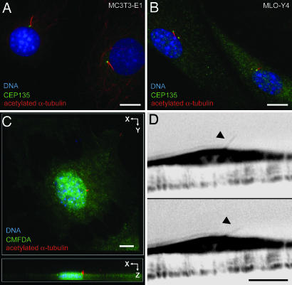
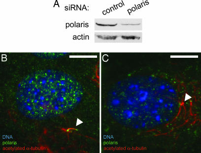
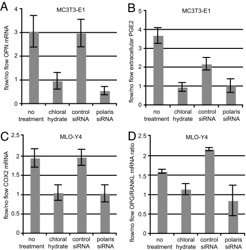
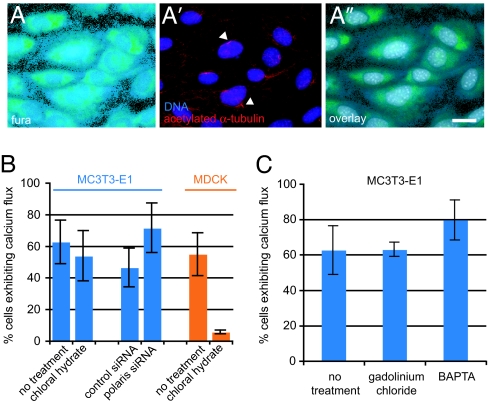
Primary cilia are sensory organelles that translate extracellular chemical and mechanical cues into cellular responses. Bone is an exquisitely mechanosensitive organ, and its homeostasis depends on the ability of bone cells to sense and respond to mechanical stimuli. One such stimulus is dynamic fluid flow, which triggers biochemical and transcriptional changes in bone cells by an unknown mechanism. Here we report that bone cells possess primary cilia that project from the cell surface and deflect during fluid flow and that these primary cilia are required for osteogenic and bone resorptive responses to dynamic fluid flow. We also show that, unlike in kidney cells, primary cilia in bone translate fluid flow into cellular responses in bone cells independently of Ca(2+) flux and stretch-activated ion channels. These results suggest that primary cilia might regulate homeostasis in diverse tissues by allowing mechanical signals to alter cellular activity via tissue-specific pathways. Our identification of a mechanism for mechanotransduction in bone could lead to therapeutic approaches for combating bone loss due to osteoporosis and disuse.


Conflict of interest statement

The authors declare no conflict of interest.

Figures

Fig. 1.
Fig. 1.
Primary cilia project from the apical surface of bone cells and bend during fluid flow. (A and B) Primary cilia stained with anti-acetylated α-tubulin (red) extend from centrosomes stained with anti-CEP135 (green) in MC3T3-E1 osteoblasts (A) and MLO-Y4 osteocytes (B). DNA is stained with DAPI (blue). (C) Maximum projection of a confocal z series shows a primary cilium marked by acetylated α-tubulin (red) extending from the apical surface marked by CMFDA (green) of an MC3T3-E1 osteoblast. DNA is stained with DAPI (blue). (D) Side view of a primary cilium (arrowheads) extending from the apical surface of an MC3T3-E1 osteoblast before (Upper) and during (Lower) application fluid flow from left to right. Images are inverted frames from SI Movie 1. (Scale bars: 5 μm.)
Fig. 2.
Fig. 2.
RNAi of polaris prevents primary cilium formation in cultured bone cells. (A) Western blot showing reduced levels of protein in MC3T3-E1 osteoblasts transfected with polaris siRNA (right lane) relative to cells transfected with control siRNA (left lane). Western blot for actin is a loading control. (B) Control MC3T3-E1 osteoblast showing polaris (green) at the primary cilium (arrowhead) marked by acetylated α-tubulin (red). DNA is stained with DAPI (blue). (C) Polaris siRNA-treated MC3T3-E1 osteoblast showing reduced polaris staining (green) and the absence of a primary cilium extending from the centrioles (arrowhead). Stable cytoplasmic microtubules are marked by acetylated α-tubulin (red) and DNA is stained with DAPI (blue). (Scale bars: 10 μm.)
Fig. 3.
Fig. 3.
Primary cilia are required for cellular responses to dynamic fluid flow in osteoblasts and osteocytes. (A) Flow-induced OPN mRNA levels in MC3T3-E1 osteoblasts with and without primary cilia. Cells were exposed to 1-Pa 1-Hz oscillatory fluid flow for 1 h, and OPN mRNA levels were quantified by real-time RT-PCR and normalized to 18S rRNA. (B) PGE2 release for MC3T3-E1 osteoblasts with and without primary cilia. Extracellular PGE2 levels were quantified by ELISA and normalized to total protein. (C) COX2 mRNA levels for MLO-Y4 osteocytes as quantified by real-time RT-PCR and normalized to18S rRNA. (D) OPG/RANKL mRNA ratios for MLO-Y4 osteocytes. [Error bars: SEM (n ≥ 6).]
Fig. 4.
Fig. 4.
Ca+2 flux during flow in bone cells is independent of primary cilia. (A) Fura 2-AM fluorescence in MC3T3-E1 osteoblasts during in vivo Ca+2 imaging. (A′) Staining of primary cilia (arrowheads) marked by acetylated α-tubulin (red) in the same field of cells after fixation; DNA is stained with DAPI (blue). (A″) Overlay of Fura 2-AM and DAPI fluorescence shows that the fields of cells are identical. (Scale bar: 10 μm.) (B) Percentage of MC3T3-E1 osteoblasts (blue bars) and MDCK cells (orange bars) exhibiting Ca+2 flux during flow (see Methods). (C) Percentage of MC3T3-E1 osteoblasts exhibiting Ca+2 flux during flow after various treatments. [Error bars: SEM (n ≥ 4).]

References

    1. Huang H, Kamm RD, Lee RT. Am J Physiol. 2004;287:C1–C11. - PubMed
    1. Hughes-Fulford M. Sci STKE. 2004;2004:RE12. - PubMed
    1. Simons M, Walz G. Kidney Int. 2006;70:854–864. - PubMed
    1. Yamamoto K, Takahashi T, Asahara T, Ohura N, Sokabe T, Kamiya A, Ando J. J Appl Physiol. 2003;95:2081–2088. - PubMed
    1. Wong M, Siegrist M, Goodwin K. Bone. 2003;33:685–693. - PubMed

Publication types

LinkOut - more resources