. 2007 Nov 15;110(10):3517-25.
doi: 10.1182/blood-2007-03-079616. Epub 2007 Aug 2.

Gambogic acid, a novel ligand for transferrin receptor, potentiates TNF-induced apoptosis through modulation of the nuclear factor-kappaB signaling pathway

Affiliations

Gambogic acid, a novel ligand for transferrin receptor, potentiates TNF-induced apoptosis through modulation of the nuclear factor-kappaB signaling pathway

Manoj K Pandey et al. Blood. .

Expression of concern in

Abstract

Gambogic acid (GA), a xanthone derived from the resin of the Garcinia hanburyi, has been recently demonstrated to bind transferrin receptor and exhibit potential anticancer effects through a signaling mechanism that is not fully understood. Because of the critical role of NF-kappaB signaling pathway, we investigated the effects of GA on NF-kappaB-mediated cellular responses and NF-kappaB-regulated gene products in human leukemia cancer cells. Treatment of cells with GA enhanced apoptosis induced by tumor necrosis factor (TNF) and chemotherapeutic agents, inhibited the expression of gene products involved in antiapoptosis (IAP1 and IAP2, Bcl-2, Bcl-x(L), and TRAF1), proliferation (cyclin D1 and c-Myc), invasion (COX-2 and MMP-9), and angiogenesis (VEGF), all of which are known to be regulated by NF-kappaB. GA suppressed NF-kappaB activation induced by various inflammatory agents and carcinogens and this, accompanied by the inhibition of TAK1/TAB1-mediated IKK activation, inhibited IkappaBalpha phosphorylation and degradation, suppressed p65 phosphorylation and nuclear translocation, and finally abrogated NF-kappaB-dependent reporter gene expression. The NF-kappaB activation induced by TNFR1, TRADD, TRAF2, NIK, TAK1/TAB1, and IKKbeta was also inhibited. The effect of GA mediated through transferrin receptor as down-regulation of the receptor by RNA interference reversed its effects on NF-kappaB and apoptosis. Overall our results demonstrate that GA inhibits NF-kappaB signaling pathway and potentiates apoptosis through its interaction with the transferrin receptor.


Figures

Figure 1
Figure 1
GA potentiates apoptotic effects of TNF and chemotherapeutic agents. (A) The chemical structure of GA. (B-D) GA enhances TNF-, 5-FU–, and doxorubicin-induced cytotoxicity. Five thousand cells were seeded in triplicate in 96-well plates. The cells were pretreated with the indicated concentrations of GA and then incubated with chemotherapeutic agents. Cell viability was then analyzed by the MTT method. (E) GA enhances TNF-induced cytotoxicity. KBM-5 cells were pretreated with indicated concentrations of GA for 4 hours and then incubated with 1 nM TNF for 24 hours. The cells were stained with a live/dead assay reagent for 30 minutes and then analyzed under a fluorescence microscope (left panel). The percentage of apoptosis was plotted as mean plus or minus SD (right panel). (F) GA enhances TNF-induced annexin V–FITC binding. (G) Effect of GA on PARP cleavage. Cells were pretreated with 1.0 μM GA for 4 hours and then incubated with 1 nM TNF for the indicated times. Whole-cell extracts were prepared and analyzed by Western blotting with an anti-PARP antibody. (H) A293 cells were transiently transfected with p65 plasmids. After 24 hours, cells were treated with 1.0 μM GA for 4 hours followed by 1 nM TNF for 24 hours. Whole-cell extracts were prepared and analyzed by Western blotting with an anti-PARP antibody.
Figure 2
Figure 2
GA inhibits TNF-induced expression of NF-κB–dependent antiapoptotic, proliferative, and metastatic proteins. (A) GA inhibits the expression of TNF-induced antiapoptotic proteins. KBM-5 cells were incubated with 1.0 μM GA for 4 hours and then treated with 1 nM TNF for the indicated times. Whole-cell extracts were prepared and analyzed by Western blotting with the indicated antibodies. (B,C) GA inhibits TNF-induced expression of c-Myc, cyclin D1, COX-2, ICAM-1, MMP-9, and VEGF. KBM-5 cells were incubated with 1.0 μM GA for 4 hours and then treated with 1 nM TNF for the indicated times. Whole-cell extracts were prepared and analyzed by Western blotting with the relevant antibodies.
Figure 3
Figure 3
GA inhibits NF-κB activation induced by different stimuli. (A) GA blocks NF-κB activation induced by TNF, okadaic acid (OA), PMA, LPS, CSC, and H2O2. Human myeloid leukemia KBM-5 cells were preincubated with 5 μM GA for 4 hours and then treated with 0.1 nM TNF for 30 minutes, 500 nM OA for 4 hours, 25 ng/mL PMA for 2 hours, 10 μg/mL LPS, and 40 μg/mL CSC and 250 μM H2O2 for 1 hour each. Nuclear extracts were analyzed for NF-κB activation by EMSA. The results shown are representative of 3 independent experiments. (B) Dose-dependent effect of GA on TNF-induced NF-κB activation. KBM-5 cells were incubated with the indicated concentrations of GA for 4 hours and treated with 0.1 nM TNF for 30 minutes. The nuclear extracts were assayed for NF-κB activation by EMSA. CV (%) indicates cell viability of the cells. The results shown are representative of 3 independent experiments. (C) Time-dependent effect of GA on TNF-induced NF-κB activation. KBM-5 cells were preincubated with 5 μM GA for the indicated times and then treated with 0.1 nM TNF for 30 minutes. The nuclear extracts were prepared and assayed for NF-κB activation by EMSA. CV (%) indicates cell viability of the cells. The results shown are representative of 3 independent experiments. (D) NF-κB induced by TNF is composed of p65 and p50 subunits. Nuclear extracts from untreated cells or cells treated with 0.1 nM TNF were incubated with the indicated antibodies, an unlabeled NF-κB oligoprobe, or a mutant oligoprobe. They were then assayed for NF-κB activation by EMSA. (E) The direct effect of GA on the NF-κB–DNA binding. Nuclear extracts were prepared from untreated cells or cells treated with 0.1 nM TNF and incubated for 30 minutes with the indicated concentrations of GA. They were then assayed for NF-κB activation by EMSA. (F) Effect of plumbagin (PG) and GA on the binding of NF-κB to DNA. Nuclear extracts were prepared from untreated cells or cells treated with 0.1 nM TNF and incubated for 30 minutes with the 5-μM concentrations of GA and PG. They were then assayed for NF-κB activation by EMSA.
Figure 4
Figure 4
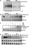
GA inhibits TNF-induced IκBα degradation, IκBα phosphorylation, and IKK activation. (A) GA inhibits TNF-induced activation of NF-κB. KBM-5 cells were incubated with 5 μM GA for 4 hours, treated with 0.1 nM TNF for the indicated times, and then analyzed for NF-κB activation by EMSA. (B) Effect of GA on TNF-induced degradation of IκBα. KBM-5 cells were incubated with 5 μM GA for 4 hours and treated with 0.1 nM TNF for the indicated times. Cytoplasmic extracts were prepared and analyzed by Western blotting with antibodies against anti-IκBα. Equal protein loading was evaluated by β-actin. (C) Effect of GA on the phosphorylation by IκBα by TNF. Cells were preincubated with 5 μM GA for 4 hours, incubated with 50 μg/mL ALLN for 30 minutes, and then treated with 0.1 nM TNF for 10 minutes. Cytoplasmic extracts were fractionated and then subjected to Western blotting with phospho-specific IκBα antibody. The same membrane was reblotted with β-actin. (D) Effect of GA on the activation of IKK by TNF. KBM-5 cells were preincubated with 5 μM GA for 4 hours and then treated with 1 nM TNF for the indicated times. Whole-cell extracts were immunoprecipitated with antibody against IKK-α and analyzed by an immune complex kinase assay. To examine the effect of GA on the level of expression of IKK proteins, whole-cell extracts were fractionated on SDS-PAGE and examined by Western blot analysis with anti–IKK-α and anti–IKK-β antibodies.
Figure 5
Figure 5
GA inhibits TNF-induced phosphorylation and nuclear translocation of p65. (A) Immunocytochemical analysis of p65 localization. KBM-5 cells were first treated with 5 μM GA for 4 hours at 37°C and then exposed to 0.1 nM TNF for 15 minutes. After the cells were centrifuged, they underwent immunocytochemical analysis. (B,C) GA inhibits TNF-induced nuclear translocation and phosphorylation of p65. KBM-5 cells were either untreated or pretreated with 5 μM GA for 4 hours at 37°C and then treated with 0.1 nM TNF for the indicated times. Nuclear extracts were prepared and analyzed by Western blotting with antibodies against p65 and phospho-specific p65. For loading control of nuclear protein, the membrane was blotted with anti-PARP antibody.
Figure 6
Figure 6
GA inhibits NF-κB activation induced by different molecules in NF-κB signaling pathway. (A) GA inhibited TNF-induced NF-κB–dependent reporter gene (SEAP) expression. A293 cells treated with the indicated concentrations of GA were transiently transfected with a NF-κB–containing plasmid linked to the SEAP gene. After 24 hours in culture with 0.1 nM TNF, cell supernatants were collected and assayed for SEAP activity. Results are expressed as fold activity over the activity of the vector control. (B,C) GA inhibited NF-κB–dependent reporter gene expression induced by TNF, TNFR1, TRADD, NIK, TRAF2, TAK1/TAB1, IKK, p65, and receptor-interacting protein (RIP). A293 cells were transiently transfected with the indicated plasmids along with a NF-κB–containing plasmid linked to the SEAP gene. Bars indicate standard deviation.
Figure 7
Figure 7
The effect of GA on NF-κB and on apoptosis are mediated through the transferrin receptor. (A) Down-regulation of TfR1 by RNA interference reverses the effect of GA. A293 cells were transfected with TfR1 si RNA or scrambled (SC) control. After 48 hours, cells were harvested and used for RNA isolation and for protein extraction. RT-PCR was done to determine the TfR1 mRNA expression. RNA levels were normalized to glyceraldehyde-3-phosphate dehydrogenase (GAPDH). Whole-cell extracts were analyzed by Western blotting with an anti-TfR1 antibody. (B) Transfected cells were preincubated with 5 μM GA for 4 hours and then treated with 0.1 nM TNF for 30 minutes. The nuclear extracts were prepared and assayed for NF-κB activation by EMSA. (C) Transfected cells were treated with 1 μM GA for 4 hours followed by 1 nM TNF for 24 hours. The cells were stained with a live/dead assay reagent and analyzed under a fluorescence microscope.

Similar articles

Cited by

References

    1. Aggarwal BB, Ichikawa H, Garodia P, et al. From traditional Ayurvedic medicine to modern medicine: identification of therapeutic targets for suppression of inflammation and cancer. Expert Opin Ther Targets. 2006;10:87–118. - PubMed
    1. Newman DJ, Cragg GM, Snader KM. Natural products as sources of new drugs over the period 1981–2002. J Nat Prod. 2003;66:1022–1037. - PubMed
    1. Guo QL, Lin SS, You QD, et al. Inhibition of human telomerase reverse transcriptase gene expression by gambogic acid in human hepatoma SMMC-7721 cells. Life Sci. 2006;78:1238–1245. - PubMed
    1. Zhang HZ, Kasibhatla S, Wang Y, et al. Discovery, characterization and SAR of gambogic acid as a potent apoptosis inducer by a HTS assay. Bioorg Med Chem. 2004;12:309–317. - PubMed
    1. Zhao L, Guo QL, You QD, Wu ZQ, Gu HY. Gambogic acid induces apoptosis and regulates expressions of Bax and Bcl-2 protein in human gastric carcinoma MGC-803 cells. Biol Pharm Bull. 2004;27:998–1003. - PubMed

Publication types

MeSH terms