. 2007 Aug 3;317(5838):663-6.
doi: 10.1126/science.1139857.

Forced unfolding of proteins within cells

Affiliations

Forced unfolding of proteins within cells

Colin P Johnson et al. Science. .

Abstract

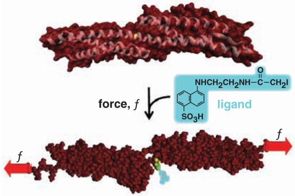
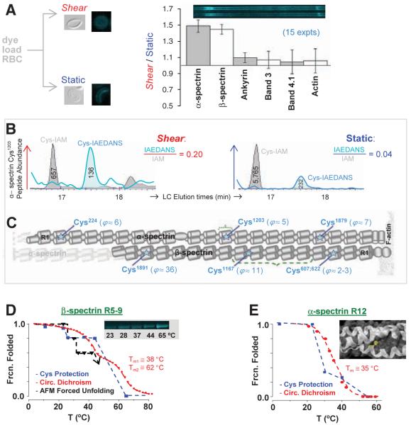
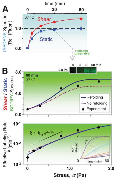
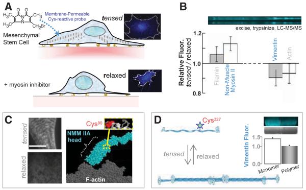
To identify cytoskeletal proteins that change conformation or assembly within stressed cells, in situ labeling of sterically shielded cysteines with fluorophores was analyzed by fluorescence imaging, quantitative mass spectrometry, and sequential two-dye labeling. Within red blood cells, shotgun labeling showed that shielded cysteines in the two isoforms of the cytoskeletal protein spectrin were increasingly labeled as a function of shear stress and time, indicative of forced unfolding of specific domains. Within mesenchymal stem cells-as a prototypical adherent cell-nonmuscle myosin IIA and vimentin are just two of the cytoskeletal proteins identified that show differential labeling in tensed versus drug-relaxed cells. Cysteine labeling of proteins within live cells can thus be used to fluorescently map out sites of molecular-scale deformation, and the results also suggest means to colocalize signaling events such as phosphorylation with forced unfolding.


Figures

Fig. 1
Fig. 1
Force-induced changes in protein structure within cells are hypothesized to expose novel binding sites for ligands. This example of a molecular dynamics simulation shows that Cys1167 in β-spectrin exposes 0 Å2 surface area (of 224 Å2) until forced extension [e.g., (15)] exposes the -SH for reaction with a thiol-reactive fluorescent dye.
Fig. 2
Fig. 2
In-cell labeling of RBCs under stressed versus static conditions demonstrates force-enhanced labeling of spectrin sites with Cys identification by quantitative MS. (A) Inset images show shear-distorted or round RBC ghosts fixed by glutaraldehyde, as well as fluorescence microscopy of membranes after IAEDANS reaction under either shear (stress σ = 0.93 Pa) or static conditions (60 min, 37°C). SDS 1D-PAGE separations of ghost lysates demonstrate shear-enhanced labeling of α-and β-spectrin, but no significant differences for other membrane proteins (±SD). (B) Extracted ion chromatograms for α12's Cys1203-containing peptide from shear and static samples. Ratios of peak areas for IAEDANS- and IAM-labeled peptides provide measures of relative dye labeling. (C) Positions along α- and β-spectrin with shear/static-labeling differentials, ϕ ≥ 2 (seetable S1). (D) Recombinant β-R5-9 construct labeled in solution by IAEDANS with increasing temperature reveals a step-wise increase in labeling extent; this is inverted and normalized to report fraction folded. The increase at T > 23°C coincides with unfolding of repeat β9, as determined by both circular dichroism (CD) and forced unfolding with an AFM. The numbered Tm are the two melting temperatures from CD. (E) Homology model of α-R12 with Cys1203 highlighted in yellow. Fraction of α12 folded versus temperature based on cysteine labeling results (blue) and CD measurements (red) (28).
Fig. 3
Fig. 3
Stress-dependent kinetics and sequential two-dye labeling for amplification. (A) Sheared (σ = 0.93 Pa) versus static RBCs show distinct labeling kinetics for spectrin. IAEDANS labeling (at 2.5 mM) of the static-labeled, surface-exposed spectrin sites fit a first-order rate of 0.13 min-1; the shear-exposed sites (66% more from fit) label more slowly at 0.04 min-1. (B) Sequential labeling entails labeling of the surface Cys (blue) for 30 min, after which BODIPY-IA (green) is added at >10-fold excess (6 mM). Half of the cells are then exposed to shear with timed aliquots again quenched by IAM and analyzed by densitometry of the green fluorescent spectrin. BODIPY-spectrin at 60 min increases with σ (top) as does the effective rate of labeling (bottom). Error bars (±SD) indicate three or more measurements (six for σ = 0). The data are fit by the exact solution of the fLL reaction for P(t), either with or without refolding (SOM text). The intermediate regime of stress (dotted black line) is dominated by unfolding and increases exponentially with stress, exhibiting a characteristic stress of σo ≈ 0.5 Pa and giving k1f ≈ 0.004 min-1. (Inset) The effect of force on computed kinetics using the fit parameters.
Fig. 4
Fig. 4
In-cell labeling of human stem cells either tensed or relaxed. (A) The membrane-permeable Cys-reactive fluorophore, mBBr, was added to 1-week cultures of MSCs with active myosins and tensed cytoskeletons and also to MSCs treated with myosin-inhibiting blebbistatin for 1 day to relax the cells. Imaging shows homogeneous labeling with 0.5 mM mBBr for 40 min. (B) SDS-PAGE and densitometry of samples (±SD, three experiments) that were either blebbistatin treated (relaxed) or untreated (tensed) show several protein bands in which the fluorescence intensities are different (normalized to protein load). Lysates were quenched with β-mercaptoethanol (50 mM) before analysis. (C) Immunofluorescence imaging of NMM IIA in tensed cells (top) and relaxed cells (bottom) indicates a spatial redistribution of myosin with drug. Scale, 5 μm. MS analyses of excised myosin bands detected labeling of Cys90, which appears buried within the fold of NMM IIA homology models. (D) Vimentin labeling in monomeric, and polymeric forms display different degrees of fluorescence (error bar from two experiments), which indicates that polymerization sterically blocks Cys327 for labeling.

References

    1. Evans EA, Skalak R. Mechanics and Thermodynamics of Biomembranes. CRC Press; Boca Raton, FL: 1980. p. 190.
    1. Erickson HP. Proc. Natl. Acad. Sci. U.S.A. 1994;91:10114. - PMC - PubMed
    1. Balaban NQ, et al. Nat. Cell Biol. 2001;3:466. - PubMed
    1. Beningo KA, Dembo M, Kaverina I, Small JV, Wang YL. J. Cell Biol. 2001;153:881. - PMC - PubMed
    1. Alenghat FJ, Ingber DE. Sci. STKE. 2002;2002(119):PE6. - PubMed

Publication types

MeSH terms