

Fundamental elements for successful performance of CT colonography (virtual colonoscopy)
- PMID: 17673837
- PMCID: PMC2627155
- DOI: 10.3348/kjr.2007.8.4.264
Fundamental elements for successful performance of CT colonography (virtual colonoscopy)
Abstract
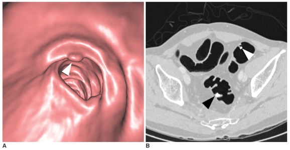
There are many factors affecting the successful performance of CT colonography (CTC). Adequate colonic cleansing and distention, the optimal CT technique and interpretation with using the newest CTC software by a trained reader will help ensure high accuracy for lesion detection. Fecal and fluid tagging may improve the diagnostic accuracy and allow for reduced bowel preparation. Automated carbon dioxide insufflation is more efficient and may be safer for colonic distention as compared to manual room air insufflation. CT scanning should use thin collimation of < or =3 mm with a reconstruction interval of < or =1.5 mm and a low radiation dose. There is not any one correct method for the interpretation of CTC; therefore, readers should be well-versed with both the primary 3D and 2D reviews. Polyps detected at CTC should be measured accurately and reported following the "polyp size-based" patient management system. The time-intensive nature of CTC and the limited resources for training radiologists appear to be the major barriers for implementing CTC in Korea.
Figures


Similar articles
-
Primary three-dimensional analysis with perspective-filet view versus primary two-dimensional analysis: evaluation of lesion detection by inexperienced readers at computed tomographic colonography in symptomatic patients.Acta Radiol. 2009 Apr;50(3):244-55. doi: 10.1080/02841850802714797. Acta Radiol. 2009. PMID: 19235581
-
Digital bowel cleansing free colonic polyp detection method for fecal tagging CT colonography.Acad Radiol. 2009 Apr;16(4):486-94. doi: 10.1016/j.acra.2008.10.011. Acad Radiol. 2009. PMID: 19268861
-
Colonic distention at CT colonography: randomized evaluation of both IV hyoscine butylbromide and automated carbon dioxide insufflation.AJR Am J Roentgenol. 2015 Jan;204(1):76-82. doi: 10.2214/AJR.14.12772. AJR Am J Roentgenol. 2015. PMID: 25539240 Clinical Trial.
-
CT colonography: techniques and applications.Radiol Clin North Am. 2009 Jan;47(1):133-45. doi: 10.1016/j.rcl.2008.11.002. Radiol Clin North Am. 2009. PMID: 19195539 Review.
-
3D detection of colonic polyps by CT colonography: accuracy, pitfalls, and solutions by adjunct 2D workup.Clin Radiol. 2015 Oct;70(10):1144-51. doi: 10.1016/j.crad.2015.07.001. Epub 2015 Jul 26. Clin Radiol. 2015. PMID: 26220124 Review.
Cited by
-
CT colonography in the diagnosis and management of colorectal cancer: emphasis on pre- and post-surgical evaluation.World J Gastroenterol. 2014 Feb 28;20(8):2014-22. doi: 10.3748/wjg.v20.i8.2014. World J Gastroenterol. 2014. PMID: 24587676 Free PMC article. Review.
-
Comparison between CT colonography and double-contrast barium enema for colonic evaluation in patients with renal insufficiency.Korean J Radiol. 2012 May-Jun;13(3):290-9. doi: 10.3348/kjr.2012.13.3.290. Epub 2012 Apr 17. Korean J Radiol. 2012. PMID: 22563266 Free PMC article.
-
Evaluation of BMI-based tube voltage selection in CT colonography: A prospective comparison of low kV versus routine 120 kV protocol.J Appl Clin Med Phys. 2023 May;24(5):e13955. doi: 10.1002/acm2.13955. Epub 2023 Mar 10. J Appl Clin Med Phys. 2023. PMID: 36897536 Free PMC article.
-
Efficacy of barium-based fecal tagging for CT colonography: a comparison between the use of high and low density barium suspensions in a Korean population - a preliminary study.Korean J Radiol. 2009 Jan-Feb;10(1):25-33. doi: 10.3348/kjr.2009.10.1.25. Korean J Radiol. 2009. PMID: 19182500 Free PMC article.
-
Pilot study on efficacy of reduced cathartic bowel preparation with polyethylene glycol and bisacodyl.World J Gastroenterol. 2013 Jan 28;19(4):561-8. doi: 10.3748/wjg.v19.i4.561. World J Gastroenterol. 2013. PMID: 23382637 Free PMC article. Clinical Trial.
References
-
- Pickhardt PJ, Choi JR, Hwang I, Butler JA, Puckett ML, Hildebrandt HA, et al. Computed tomographic virtual colonoscopy to screen for colorectal neoplasia in asymptomatic adults. N Engl J Med. 2003;349:2191–2200. - PubMed
-
- Cotton PB, Durkalski VL, Pineau BC, Palesch YY, Mauldin PD, Hoffman B, et al. Computed tomographic colonography (virtual colonoscopy): a multicenter comparison with standard colonoscopy for detection of colorectal neoplasia. JAMA. 2004;291:1713–1719. - PubMed
-
- Rockey DC, Paulson E, Niedzwiecki D, Davis W, Bosworth HB, Sanders L, et al. Analysis of air contrast barium enema, computed tomographic colonography, and colonoscopy: prospective comparison. Lancet. 2005;365:305–311. - PubMed
-
- Mulhall BP, Veerappan GR, Jackson JL. Meta-analysis: computed tomographic colonography. Ann Intern Med. 2005;142:635–650. - PubMed
-
- Ferrucci JT. Colonoscopy: virtual and optical-another look, another view. Radiology. 2005;235:13–16. - PubMed
Publication types
MeSH terms
Substances
LinkOut - more resources
Full Text Sources
Other Literature Sources
Medical
