

Case Reports
doi: 10.3348/kjr.2007.8.4.343.
Carcinosarcoma of the liver: a case report
Affiliations
- PMID: 17673846
- PMCID: PMC2627156
- DOI: 10.3348/kjr.2007.8.4.343
Item in Clipboard
Case Reports
Carcinosarcoma of the liver: a case report
Korean J Radiol.
2007 Jul-Aug.
Abstract
Primary hepatic carcinosarcoma is a rare tumor comprised of a mixture of carcinomatous and sarcomatous elements. Less than 20 adequately documented cases have been reported, however the imaging features of two cases were briefly described. We present here a case of carcinosarcoma of the liver in a 46-year-old woman, which was confirmed based on pathology. Imaging showed a large mass with large necrotic portions, small cystic portions, calcifications and bone formations.
Figures
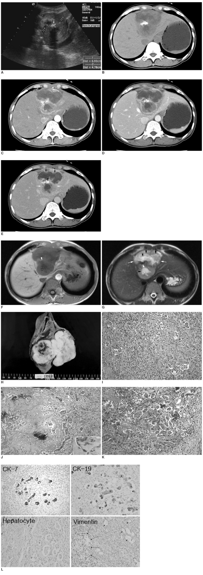
A. Longitudinal ultrasonography through the left liver showed a lobulate heterogeneous echogenic solid mass (cursors) with a highly echogenic lesion (arrow) that had posterior acoustic shadowing within it. B-E. Computed tomographic scan of this mass revealed peripheral enhancing viable portions (white arrows), large internal non-enhancing necrotic portions (black arrows), and a dense radiopaque lesion (arrowhead). The enhancing portion of this mass is hypoattenuating in precontrast scan (B), hyperattenuating in arterial phase (C), hypoattenuating in portal phase (D), and isoattenuating in equilibrium phase (E). F-G. Upon magnetic resonance imaging this mass is hypointense on the T1-weighted image (F) and hyperintense on the T2-weighted image (G). On the T2-weighted images, peripheral viable portions (white arrows) are slightly hyperintense, necrotic portions (black arrows) are moderately hyperintense, and small cystic portions (black arrowheads) are very hyperintense. Calcification and ossification have low or dark signal intensity (white arrowheads) on T1- and T2-weighted images. H. A section of gross specimen showing a well-demarcated, dumbbell-shaped, multilobulate, grayish white, solid and firm mass with a central area of necrosis and hemorrhage, measuring 8.0 cm in its greatest dimension. I. The tumor mass was composed of diffusely proliferating spindle-shaped atypical cells and scattered epithelial cell clusters (Hematoxylin & Eosin staining, × 200). J. Large atypical pyknotic cells embedded in a hyaline chondroid matrix were detected mixed with adjacent anaplastic tubular epithelial clusters and anaplastic spindle cells (Hematoxylin & Eosin staining, × 200) (Inset). Calcification within the cartilage component was occasionally detected (Hematoxylin & Eosin staining, × 100). K. Transitional features of atypical spindle and epithelial cells are seen with an irregular anastomosing reddish osteoid matrix (Hematoxylin & Eosin staining, × 200). L. The epithelial component was positive for cytokeratin 7 and 19 (cholangiocytic differentiation markers), but negative for hepatocyte (hepatocytic differentiation marker). Atypical spindle cells and cartilaginous components (large arrow) were all positive for vimentin (mesenchymal differentiation marker), however the scattered epithelial clusters were negative for vimentin (small arrows).
References
-
- Freeman AJ, Bullpitt P, Keogh GW. Primary hepatic carcinosarcoma. ANZ J Surg. 2004;74:1021–1023. - PubMed
-
- Rummeny E, Weissleder R, Stark DD, Saini S, Compton CC, Bennett W, et al. Primary liver tumors: diagnosis by MR imaging. AJR Am J Roentgenol. 1989;152:63–72. - PubMed
-
- Nomura K, Aizawa S, Ushigome S. Carcinosarcoma of the liver. Arch Pathol Lab Med. 2000;124:888–890. - PubMed
-
- Wang XW, Liang P, Li HY. Primary hepatic carcinosarcoma: a case report. Chin Med J. 2004;117:1586–1587. - PubMed
Publication types
MeSH terms
Substances
LinkOut - more resources
Full Text Sources
Medical
