. 2007 Sep 5;26(17):3957-67.
doi: 10.1038/sj.emboj.7601818. Epub 2007 Aug 2.

TGF-beta activates Erk MAP kinase signalling through direct phosphorylation of ShcA

Affiliations

TGF-beta activates Erk MAP kinase signalling through direct phosphorylation of ShcA

Matt K Lee et al. EMBO J. .

Abstract

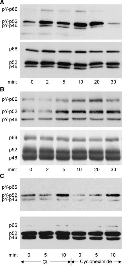
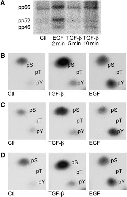
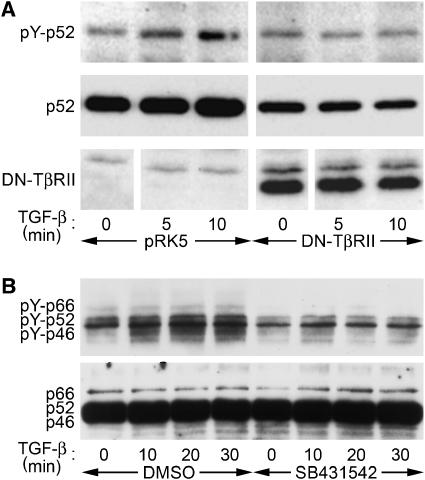
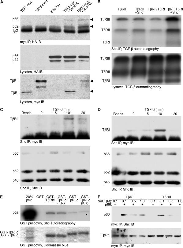
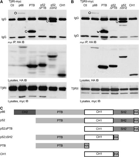
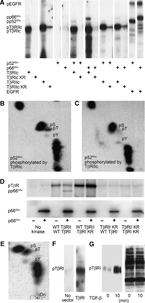
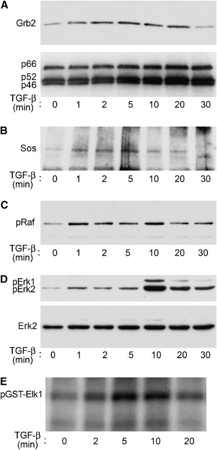
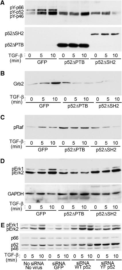
Erk1/Erk2 MAP kinases are key regulators of cell behaviour and their activation is generally associated with tyrosine kinase signalling. However, TGF-beta stimulation also activates Erk MAP kinases through an undefined mechanism, albeit to a much lower level than receptor tyrosine kinase stimulation. We report that upon TGF-beta stimulation, the activated TGF-beta type I receptor (TbetaRI) recruits and directly phosphorylates ShcA proteins on tyrosine and serine. This dual phosphorylation results from an intrinsic TbetaRI tyrosine kinase activity that complements its well-defined serine-threonine kinase function. TGF-beta-induced ShcA phosphorylation induces ShcA association with Grb2 and Sos, thereby initiating the well-characterised pathway linking receptor tyrosine kinases with Erk MAP kinases. We also found that TbetaRI is tyrosine phosphorylated in response to TGF-beta. Thus, TbetaRI, like the TGF-beta type II receptor, is a dual-specificity kinase. Recruitment of tyrosine kinase signalling pathways may account for aspects of TGF-beta biology that are independent of Smad signalling.


Figures

Figure 1
Figure 1
TGF-β induces ShcA tyrosine phosphorylation. (A, B) Anti-phosphotyrosine Western blot of ShcA immunoprecipitated from lysates of Mv1Lu cells (A) or 3T3-Swiss cells (B) treated with 4 ng/ml TGF-β for the indicated times (upper panels), together with ShcA immunoblots of the same membranes (lower panels). (C) Anti-phosphotyrosine Western blot of ShcA immunoprecipitated from lysed 3T3-Swiss cells treated with or without cycloheximide before and concomitant with stimulation with 4 ng/ml TGF-β for the indicated times. The same membrane was reprobed for ShcA (lower panel).
Figure 2
Figure 2
TGF-β induces ShcA phosphorylation on serine and tyrosine. (A) Autoradiogram of 3T3-Swiss cells cultured in the presence of 32P-[PO4] and treated with 4 ng/ml TGF-β, 20 ng/ml EGF, or neither (Ctl) for the indicated times. (BD) Phosphoamino acid analysis of in vivo 32P-phosphorylated p66ShcA (B), p52ShcA (C), or p46ShcA (D) isolated from the membrane shown in (A). The 32P-labelled amino acids migrated in the same positions as unlabelled phosphoserine and phosphotyrosine added to the reaction mixture. No 32P-labelled phosphothreonine was detected.
Figure 3
Figure 3
TβRII and TβRI receptors are required for TGF-β-induced ShcA tyrosine phosphorylation. (A) Anti-phosphotyrosine Western blot of HA-tagged p52ShcA immunoprecipitated from Mv1Lu cells that do or do not coexpress a cytoplasmically truncated, dominant-negative version of TβRII. (B) Anti-phosphotyrosine blot of ShcA immunoprecipitated from 3T3-Swiss cells treated with 4 ng/ml TGF-β for the indicated times after 30 min pretreatment with 10 μM TβRI inhibitr SB431542 or DMSO solvent.
Figure 4
Figure 4
ShcA interacts with TGF-β receptors. (A) p66ShcA and p52ShcA co-precipitate with TGF-β receptors in COS cells. Cells expressing myc-tagged TβRI or TβRII, and/or HA-tagged p66ShcA and p52ShcA, as indicated, were subjected to myc immunoprecipitation followed by HA immunoblotting (top). Expression of ShcA and TGF-β receptors was monitored by HA (middle) or myc (bottom) Western analysis of cell lysates. (B) ShcA interacts with cell surface TGF-β receptors. 3T3-Swiss cells were transfected to express TβRII and/or TβRI with or without p66ShcA, and cell surface receptors were radiolabelled with 125I-TGF-β1 followed by chemical crosslinking. Immunoprecipitation of both endogenous and transfected ShcA followed by autoradiography (top panel) demonstrated co-precipitation of 125I-labelled TGF-β receptors with ShcA. 125I-labelled TGF-β receptors did not precipitate when the ShcA antibody was replaced with non-immune IgG (data not shown). Autoradiography of total cell lysates (below) showed equivalent levels of 125I-labelled TGF-β receptors. (C, D) Ligand-dependent interaction of endogenous ShcA and TGF-β receptors in mammalian cells. 3T3-Swiss cells were treated with 4 ng/ml TGF-β for the indicated times, lysed, and subjected to ShcA immunoprecipitation followed by TβRII (C) or TβRI (D) immunoblotting. Control precipitations without primary antibody are shown (‘beads'). Subsequent ShcA immunoblots (lower panels) confirmed equivalent precipitation efficiencies. (E) In vitro association of p52ShcA with TβRI and TβRII cytoplasmic domains. In vitro translated 35S-labelled p52ShcA was incubated with GST, GST-fused TβRI or TβRII cytoplasmic domains, or their kinase-deficient point mutants (TβRIc, TβRIIc, TβRIc KR, and TβRIIc KR, respectively). 35S-labelled ShcA adsorbed to the GST fusion proteins was visualised by SDS–PAGE and autoradiography (upper panel). To confirm identity, 35S-labelled p52ShcA was loaded in lane 1 at 20% of the volume used in the binding assay. Equivalent GST fusion protein loading was confirmed by Coomassie blue staining. (F) ShcA association with TβRI and TβRII cytoplasmic domains in vitro depends on NaCl concentration. Myc-tagged proteins incorporating either TβRIc or TβRIIc were expressed in COS cells, isolated by myc immunoprecipitation, and bound to recombinant p66ShcA. The complexes were washed in otherwise identical buffers containing the indicated NaCl concentrations and subjected to ShcA immunoblotting (above). Myc Western analysis of the blot confirmed equivalent recovery of TβRIc and TβRIIc (below).
Figure 5
Figure 5
ShcA associates with TGF-β receptors through its PTB domain. p66ShcA differs from p52ShcA only by an N-terminal addition of a CH2 domain; otherwise, both p66ShcA and p52ShcA are comprised of PTB and SH2 phosphotyrosine-binding domains flanking a central CH1 domain. HA-tagged p66ShcA, ShcA fragments, or p52ShcA deficient in the SH2 domain (ΔSH2) or the PTB domain (ΔPTB) were expressed in 293 cells together with myc-tagged cytoplasmic domains of TβRI or TβRII. (A, B) HA Western blot of myc immunoprecipitates (top), HA immunoblot (centre), and myc immunoblot (bottom) of cells co-transfected with TβRI-myc (A) or TβRII-myc (B) and ShcA constructs. ‘○' indicates ShcA construct co-precipitated with TGF-β receptor. (C) Schematic representation of ShcA expression constructs used.
Figure 6
Figure 6
TGF-β receptors directly phosphorylate ShcA. (A) ShcA phosphorylation by purified GST-fused TβRI or TβRII cytoplasmic domains in vitro. Recombinant p52ShcA or p66ShcA was incubated with GST-fused wild type and/or kinase-inactive mutants (TβRIc, TβRIc KR, TβRIIc, or TβRIIc KR) or human EGF receptor in the presence of γ-32P-ATP. The radiolabelled, autophosphorylated cytoplasmic domains, and ShcA proteins are indicated. (B, C) Phosphoamino acid analyses of p52ShcA phosphorylated by TβRI (B) or TβRII (C) cytoplasmic domains, as shown in panel (A). The locations of radiolabelled phosphoserine (pS), phosphotyrosine (pY), and phosphothreonine (pT) correlated with those of unlabelled phosphoamino acids added to the reaction mixture. (D) In vitro phosphorylation of E. coli-derived p66ShcA by TβRII-TβRI cytoplasmic chimeras generated in insect cells. Wild-type (WT) or kinase-inactivated (KR) receptor cytoplasmic domains were incubated with or without p66ShcA. The radiolabelled p66ShcA and autophosphorylated RII-RI chimeras are visualised in the upper panel. ShcA immunoblotting confirmed equivalent p66ShcA levels in the reaction mixtures (lower panel). (E) Phosphoamino acid analyses of p66ShcA phosphorylated by wtRII-wtRI, as shown in panel (D). (F) Phosphotyrosine immunoblot of in vitro kinase reaction products containing myc immunoprecipitates from COS cells transfected with control pRK5 plasmid or pRK5 encoding myc-tagged TβRI. (G) TGF-β induces tyrosine phosphorylation of endogenous TβRI, as assessed by TβRI immunoblot of anti-pTyr immunoprecipitates from Mv1Lu cells treated with TGF-β for 10 min (left). Equal precipitation efficiency was demonstrated by phosphotyrosine immunoblotting of the precipitates (right).
Figure 7
Figure 7
TGF-β activates the ShcA pathway in 3T3-Swiss cells. (A) TGF-β induces ShcA to associate with Grb2. Cells were stimulated with TGF-β for the indicated times, lysed, and subjected to ShcA immunoprecipitation followed by Grb2 Western analysis (upper panel). Reprobing the same membrane for ShcA confirmed equivalent precipitation efficiency (lower panel). (B) TGF-β induces ShcA to associate with Sos. The upper portion of the membrane shown in (A) was immunoblotted using an anti-Sos antibody. (C) TGF-β induces c-Raf phosphorylation, as assessed by phosphoRaf immunoblotting of stimulated cell lysates. (D) TGF-β induces Erk1/2 phosphorylation. The membrane in (C) was immunoblotted using a phosphoErk1/2-specific antibody (above). Reprobing the blot for Erk2 showed equivalent Erk expression and loading (below). (E) TGF-β activates Erk1/2. Stimulated cell lysates were incubated in vitro with Elk1-GST and γ32P-ATP; 32P-labelled Elk1-GST was visualised by autoradiography.
Figure 8
Figure 8
Erk activation by TGF-β is inhibited by inactive ShcA mutants and ShcA downregulation. 3T3-Swiss cells were infected with adenoviruses expressing GFP (control), HA-tagged p52ShcA lacking the PTB domain (p52ShcAΔPTB), or HA-tagged p52ShcA lacking the SH2 domain (p52ShcAΔSH2). Cells were then stimulated with TGF-β for the indicated times and lysed. (A) TGF-β-induced tyrosine phosphorylation of endogenous ShcA is attenuated by expression of p52ShcA truncations. Endogenous ShcA was immunoprecipitated and subjected to phosphotyrosine Western analysis (above). HA immunoblotting of this membrane confirmed expression of p52ShcA truncations and equivalent precipitation efficiency (below). (B) TGF-β-induced Grb2 association with endogenous ShcA is attenuated by p52ShcA truncations. The membrane in (A) was subjected to Grb2 Western analysis. (C) TGF-β-induced Raf phosphorylation is attenuated by p52ShcA truncations. Cell lysates from the experiment shown in (A) were subjected to phosphoRaf Western blot. (D) TGF-β-induced Erk phosphorylation is attenuated by p52ShcA truncations. The membrane shown in (C) was subjected to phosphoErk1/2 Western blot (above). The membrane was reprobed for GAPDH to confirm equivalent loading (below). (E) TGF-β-induced Erk activation is attenuated by ShcA silencing and restored by ectopic expression of wild-type, but not mutant p52ShcA. 3T3-Swiss cells were exposed to an siRNA targeted to the common region of ShcA together with adenoviruses encoding either green fluorescent protein (GFP), wild-type p52ShcA, or p52ShcA in which the Grb2 binding sites are mutated. Both p52ShcA constructs incorporate a silent mutation in the siRNA target site. PhosphoErk immunoblot of cells stimulated with TGF-β for the indicated times (above). ShcA immunoblot confirming partial downregulation and virus-mediated expression (below).

References

    1. Attisano L, Wrana JL (2002) Signal transduction by the TGF-β superfamily. Science 296: 1646–1647 - PubMed
    1. Berrou E, Fontenay-Roupie M, Quarck R, McKenzie FR, Lévy-Toldano S, Tobelem G, Bryckaert M (1996) Transforming growth factor β1 inhibits mitogen-activated protein kinase induced by basic fibroblast growth factor in smooth muscle cells. Biochem J 316: 167–173 - PMC - PubMed
    1. Cano E, Hazzalin CA, Kardalinou E, Buckle RS, Mahadevan LC (1995) Neither ERK nor JNK/SAPK MAP kinase subtypes are essential for histone H3/HMG-14 phosphorylation or c-fos and c-jun induction. J Cell Sci 108: 3599–3609 - PubMed
    1. Chen RH, Moses HL, Maruoka EM, Derynck R, Kawabata M (1995) Phosphorylation-dependent interaction of the cytoplasmic domains of the type I and type II transforming growth factor-β receptors. J Biol Chem 270: 12235–12241 - PubMed
    1. Cooper JA, Sefton BM, Hunter T (1983) Detection and quantification of phosphotyrosine in proteins. Methods Enzymol 99: 387–402 - PubMed

Publication types

MeSH terms

Substances