. 2007 Aug 22;26(16):3794-803.
doi: 10.1038/sj.emboj.7601811. Epub 2007 Aug 2.

J-protein co-chaperone Sis1 required for generation of [RNQ+] seeds necessary for prion propagation

Affiliations

J-protein co-chaperone Sis1 required for generation of [RNQ+] seeds necessary for prion propagation

Rebecca Aron et al. EMBO J. .

Abstract

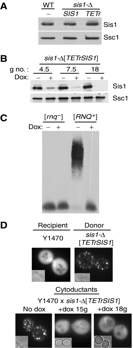
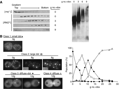
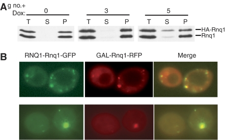
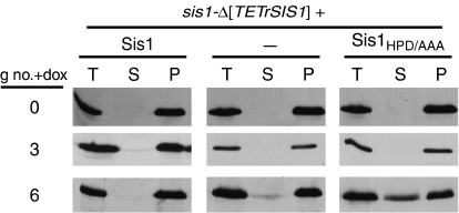
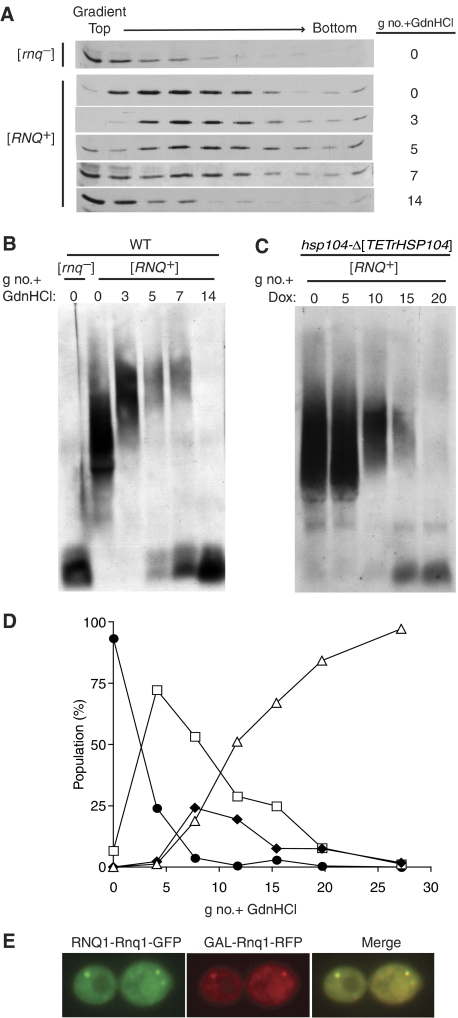
Yeast prions are protein-based genetic elements capable of self-perpetuation. One such prion, [RNQ(+)], requires the J-protein Sis1, an Ssa Hsp70 co-chaperone, as well as the AAA+ ATPase, Hsp104, for its propagation. We report that, upon depletion of Sis1, as well as upon inactivation of Hsp104, Rnq1 aggregates increased in size. Subsequently, cells having large aggregates, as well as an apparently soluble pool of Rnq1, became predominant in the cell population. Newly synthesized Rnq1 localized to both aggregates and bulk cytosol, suggesting that nascent Rnq1 partitioned into pools of prion and nonprion conformations, and implying that these large aggregates were still active as seeds. Ultimately, soluble Rnq1 predominated, and the prion was lost from the population. Our data suggest a model in which J-protein:Hsp70 machinery functions in prion propagation, in conjunction with Hsp104. Together, these chaperones facilitate fragmentation of prion polymers, generating a sufficient number of seeds to allow efficient conversion of newly synthesized Rnq1 into the prion conformation.


Figures

Figure 1
Figure 1
Loss of [RNQ+] upon reduction of Sis1 levels. (A) Expression of Sis1 from the TETr promoter. Extracts were prepared from wild-type (wt) cells of sis1-Δ cells expressing Sis1 under the control of either its native promoter (SIS1) or the TETr promoter (TETr). Protein was separated by SDS–PAGE and subjected to immunoblot analysis using antibodies specific for Sis1 or, as a loading control, Ssc1. (B) sis1-Δ[TETrSIS1] cells cultured for the indicated number of generations (g no.) in the absence (−) or presence (+) of doxycycline (dox). Lysates were prepared and subjected to SDS–PAGE and immunoblot analysis using antibodies specific for Sis1 or, as a loading control, Ssc1. (C, D) Effect of reduced SIS1 expression by repression of the TETr promoter. (C) sis1-Δ[TETrSIS1] [rnq] and [RNQ+] strains subcultured in the absence (−) or presence (+) of doxycycline were harvested after 16 generations. Prepared lysates were resolved by SDD–AGE and immunoblot analysis with α-Rnq1 antibodies. (D) Rnq1-GFP visualization by fluorescent microscopy of cytoduction recipient (Y1470) and donor sis1-Δ[TETrSIS1]) strains transiently overexpressing CUP1-Rnq1-GFP (top). sis1-Δ[TETrSIS1] cells subcultured in the absence (no dox) or presence of doxycycline (+dox) for up to 15–18 generations (g) cytoduced with Y1470, and transiently overexpressing CUP1-Rnq1-GFP, visualized by fluorescence microscopy (bottom). In each case, eight or more individual cytoductants were analyzed and >200 cells were observed; >99% of cells in each sample showed either diffuse or punctate fluorescence. Representative cells are shown.
Figure 2
Figure 2
SIS1 repression alters state of [RNQ+] before curing cells of prion. (A) Lysates from sis1-Δ[TETrSIS1] [rnq] or [RNQ+] cells treated with doxycycline for the indicated number of generations (g no.+dox) were (left panel) separated by centrifugation through sucrose gradients, and fractions were isolated and analyzed by SDS–PAGE and immunoblotted with α-Rnq1, or (right panel) resolved by SDD–AGE and immunoblot analysis with α-Rnq1. (B) Cells expressing Rnq1-GFP from the RNQ1 promoter were visualized by fluorescence microscopy. Representative images of classes Rnq1-GFP phenotypes: small dots (•), large dot (□), diffuse+dot (⧫), diffuse (▵) (left panel). The percentage of cells that exhibited the indicated Rnq1-GFP phenotype, out of >800 cells per sample, plotted against the number of generations cells were subcultured in doxycycline (g no.+dox) (right panel). See Supplementary Table 1.
Figure 3
Figure 3
Accumulation of newly synthesized Rnq1 after SIS1 repression. (A) sis1-Δ[TETrSIS1] cells transformed with GAL-HA-Rnq1 were subcultured in rich media in the presence of doxycycline for 0, 3 or 5 generations (g no.+dox). Cells were harvested, washed and transferred to doxycycline-containing rich, galactose-based media to induce HA-Rnq1 expression. After 3 h in galactose, extracts were prepared and subjected to high-speed centrifugation. Total (T), supernatant (S) and pellet (P) fractions were resolved by SDS–PAGE and analyzed by immunoblotting with α-Rnq1. (B) sis1-Δ[TETrSIS1] cells constitutively expressing Rnq1-GFP and harboring the GAL-Rnq1-RFP plasmid were cultured in doxycycline-containing glucose-based medium for 8 (upper panel) or 12 (lower panel) generations, and then cultured in the galactose-based medium in the presence of doxycycline for 3 h. Shown are two examples of diffuse+dot phenotype filtered for GFP, RFP or the overlay of the two images with yellow indicating colocalization.
Figure 4
Figure 4
Sis1 is an Hsp70 co-chaperone in prion maintenance. sis1-Δ[TETrSIS1] cells expressing wild-type Sis1, empty vector (−), or Sis1HPD/AAA were cultured in the presence of doxycycline for the indicated number of generations (g no.+dox). Total (T), supernatant (S) and pellet (P) fractions of lysates following centrifugation were resolved by SDS–PAGE and immunoblot analysis with α-Rnq1.
Figure 5
Figure 5
Hsp104 and Sis1 inhibition result in similar pattern of distribution of Rnq1 over time. (A, B) GdnHCl treatment. Lysates prepared from wild-type [rnq] and [RNQ+] cells subcultured in the presence of GdnHCl for 0, 3, 5, 7 or 14 generations (g no.) were separated by (A) sucrose gradient centrifugation, followed by SDS–PAGE or (B) SDD–AGE. Rnq1 was detected by immunoblotting with α-Rnq1 antibodies. (C) Depletion of Hsp104. Lysates prepared from hsp104-Δ[TETrHSP104] cells treated with 10 μg/ml of doxycycline were harvested 0, 5,10, 15 and 20 generations (g no.) were detected by immunoblotting with α-Rnq1 antibodies. (D) Time course of GdnHCl treatment of wild-type cells carrying the RNQ1-Rnq1-GFP plasmid. Cells were harvested after the indicated number of generations (g no.+GdnHCl), and visualized by fluorescence microscopy. Plots represent the percentage of cells, out of >500 per sample, that exhibited the indicated Rnq1-GFP classification (small dots (•), large dot (□), diffuse+dot (⧫), diffuse (▵)) plotted against number of generations cells were treated with GdnHCl (g no.+GdnHCl). See Supplementary Table 2. (E) Wild-type cells constitutively expressing Rnq1-GFP from the RNQ1 promoter and carrying the plasmid capable of expressing Rnq1-RFP in galactose-based medium were grown in glucose-based medium in the presence of GdnHCl for eight generations, then washed and resuspended in galactose-based medium for an additional 3 h. Shown is an example of diffuse+dot phenotype filtered for GFP, RFP or the overlay of the two images with yellow indicating colocalization.

References

    1. Bagriantsev S, Liebman SW (2004) Specificity of prion assembly in vivo. [PSI+] and [PIN+] form separate structures in yeast. J Biol Chem 279: 51042–51048 - PubMed
    1. Borchsenius AS, Muller S, Newnam GP, Inge-Vechtomov SG, Chernoff YO (2006) Prion variant maintained only at high levels of the Hsp104 disaggregase. Curr Genet 49: 21–29 - PubMed
    1. Bosl B, Grimminger V, Walter S (2006) The molecular chaperone Hsp104—a molecular machine for protein disaggregation. J Struct Biol 156: 139–148 - PubMed
    1. Brachmann A, Baxa U, Wickner RB (2005) Prion generation in vitro: amyloid of Ure2p is infectious. EMBO J 24: 3082–3092 - PMC - PubMed
    1. Bradley ME, Bagriantsev S, Vishveshwara N, Liebman SW (2003) Guanidine reduces stop codon read-through caused by missense mutations in SUP35 or SUP45. Yeast 20: 625–632 - PubMed

Publication types

MeSH terms