. 1976 Apr;142(4):487-505.

Orthotopic liver transplantation in ninety-three patients

Orthotopic liver transplantation in ninety-three patients

T E Starzl et al. Surg Gynecol Obstet. 1976 Apr.

Abstract

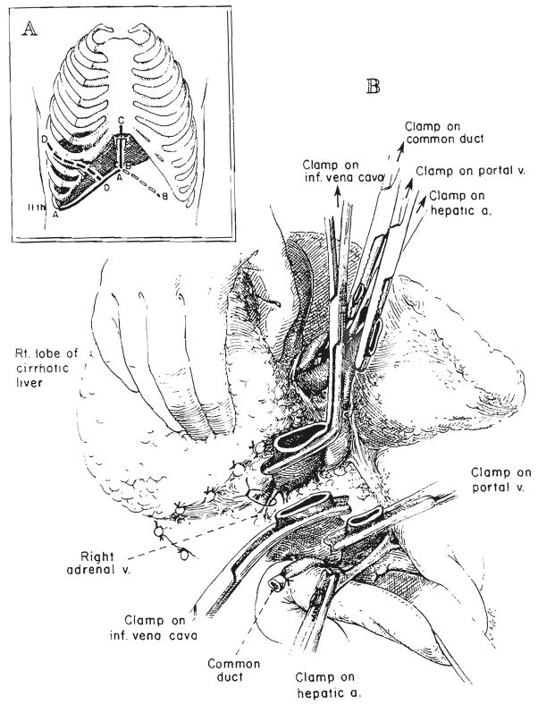
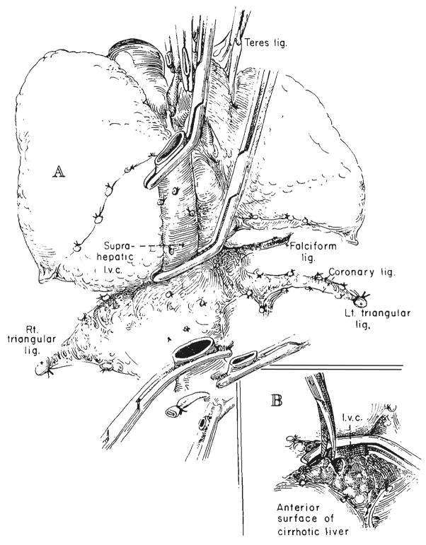
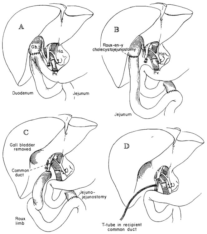
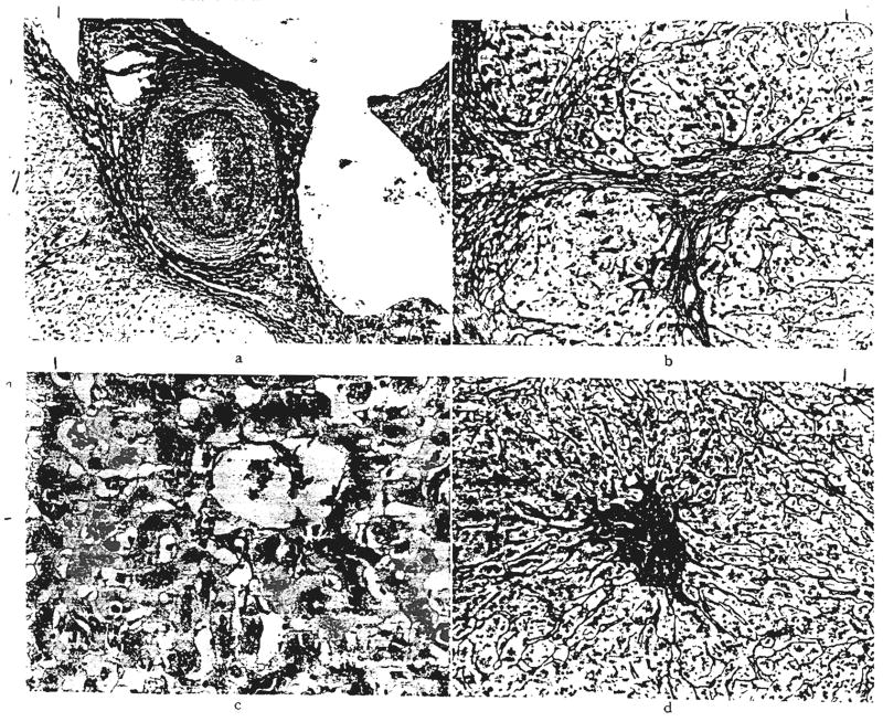
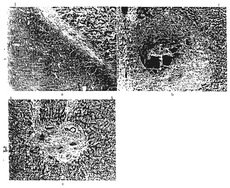
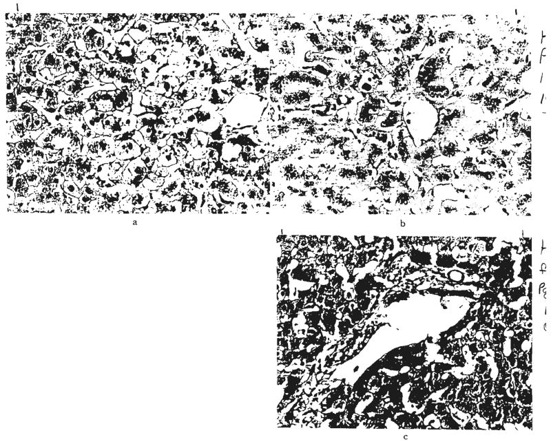
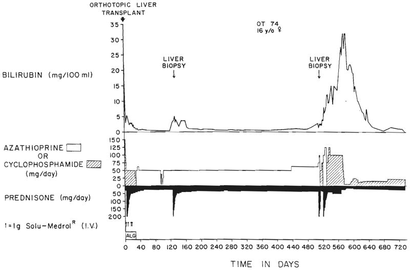
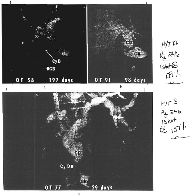
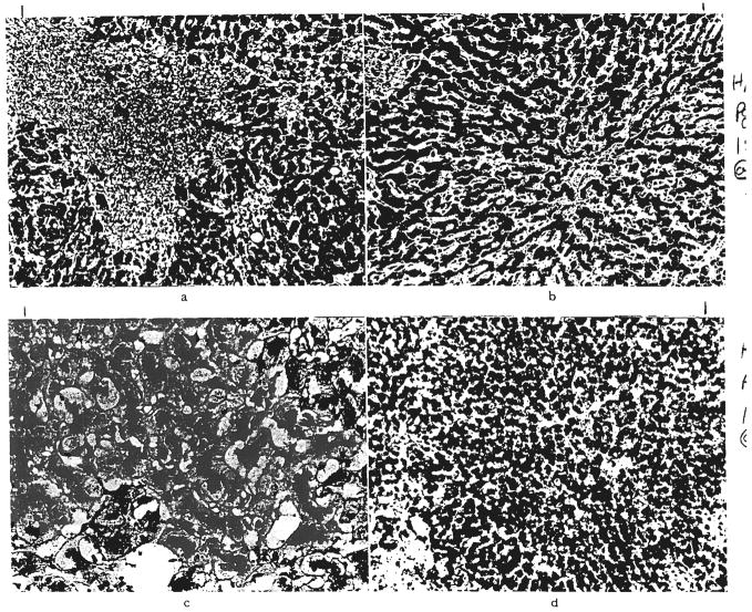
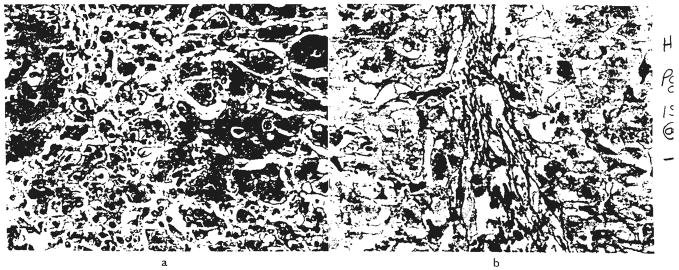
During the 11 1/2 year period ending 13 months ago, 93 consecutive patients were treated with orthotopic liver transplantation. Fifty-six of the recipients were 18 years old or younger, and the other 37 were adults. The most common indications for operation were biliary atresia, primary hepatic malignant tumor, chronic aggressive hepatitis and alcoholic cirrhosis. There has been a gradual improvement in results throughout the period of study, although to a satisfactory level. Twenty-seven of the 93 patients survived for at least one year after liver replacement with a maximum of six years, and 16 are still alive after 13 to 71 months. The 11 late deaths after one to six years were caused by chronic rejection, biliary obstruction, recurrence of hepatoma, systemic infection or hepatitis of the homograft. Rejection of the liver as judged by classical histopathologic criteria played a surprisingly small role in the heavy over-all mortality, accounting for less than 10 per cent of the deaths. Technical or mechanical problems, especially those of biliary duct reconstruction, were a far greater cause of failure, as were systemic infections. Six of the 37 adult recipients had lethal cerebrovascular accidents during, or just after, operation. When abnormalities of liver function developed in the postoperative period, the nearly automatic diagnosis of homograft rejection, in retrospect, proved to have been wrong in most instances. Further development of liver transplantation depends upon two kinds of progress. There must be reduction of operative and early postoperative accidents and complications by more discriminating patient selection, purely technical improvement and better standardization of biliary duct reconstruction. The second area will be in sharpening the criteria for the differnetial diagnosis of postoperative hepatic malfunction, including the liberal use of transhepatic cholangiography and needle biopsy. Only then can better decisions be made about changes in medication or about the need for secondary corrective surgical procedures.


Figures

Fig. 1
Fig. 1
Technique of retrograde removal of liver. A, Incisions. AA, Subcostal incision used for all orthotopic liver transplantations. BB, CC and DD, Frequently used extensions from the AA incision. B. Beginning retrograde removal after transection of inferior vena cava and hilar structures. All posterior tissue that is cut should be ligated, although the named vessels encountered, such as the right adrenal vein, are few in number.
Fig. 2
Fig. 2
A, Operative field after retrograde liver mobilization. The last remaining structure, the suprahepatic inferior vena cava, has been clamped above the liver. B, Technique for mobilizing a suitable length of suprahepatic vena cava after placement of clamp. In adults, this usually involves cutting away cirrhotic liver tissue over the frequently distorted and foreshortened right and left hepatic veins.
Fig. 3
Fig. 3
Techniques of biliary duct reconstruction used for most of the transplantation recipients. A, Cholecystoduodenostomy. B, Cholecystojejunostomy. C, Choledochojejunostomy after removal of gallbladder. D, Choledochocholedochostomy. Note that the T-tube is placed, if possible, in recipient common duct.
Fig. 4
Fig. 4
Acute rejection. Hepatic homocraft, OT 31, nine days after transplantation. Large numbers of lymphoid cells are present in the portal tracts and between the damaged hepatocytes. Hematoxylin and eosin, a, ×300; b, ×500.
Fig. 5
Fig. 5
Chronic rejection. a, A small hepatic artery branch in a graft, OT 16b, 339 days after retransplantation is almost occluded by intimal thickening. Elastic stain, ×60. b, Residual scarring after rejection. Same graft, OT 16b. There is increased portal fibrous tissue and connective tissue septums extend into the lobules. Reticulin, ×60. c, Centrilobular cholestasis with bile “thrombi” in dilated canaliculi in graft, OT 14a, one year and 16 days after transplantation. Bile stain, ×200. d, Collapse of reticulin fiber framework around central veins in graft, OT 10, 186 days after transplantation. Reticulin, ×60.
Fig. 6
Fig. 6
Large bile duct obstruction. a, Biliary cast syndrome in graft, OT 55, two years and two months after transplantation. The wall of the distended bile duct is fibrous, and the lumen is blocked by inspissated bile and debris. Hematoxylin and eosin, ×60. b, Biliary cast syndrome in graft, OT 43, 47 days after transplantation. A portal tract is greatly expanded and an intrahepatic bile duct is blocked by inspissated bile. Hematoxylin and eosin, ×50. c, Chronic large duct biliary obstruction. Enlarged fibrous portal tract with proliferated bile ductules containing bile casts. Hematoxylin and eosin, ×70.
Fig. 7
Fig. 7
Presumed drug toxicity. Biopsy of a graft in a patient, OT 74, who was deeply jaundiced at one year and five months after transplantation. The liver functions returned to normal after azathioprine was replaced with cyclophosphamide and after the prednisone dosage was lowered to less than half of its previous level. a, Centrilobular hepatocytes are swollen and show abnormal variation in nuclear size. Hematoxylin and eosin, ×200. b, Centrilobular cholestasis with bile thrombi in dilated canaliculi. Bile stain, ×200. c, Normal portal tract. Hematoxylin and eosin, ×200.
Fig. 8
Fig. 8
The first two post-transplantation years of patient, OT 74. The second liver biopsy, which is shown in Figure 7, suggested drug toxicity. Substitution of cyclophosphamide for azathioprine and reductions in steroid dosage were followed by gradual resolution of the profound hyperbilirubinemia.
Fig. 9
Fig. 9
Examples revealed by transhepatic cholangiography of homograft cystic duct obstruction after biliary reconstruction with cholecystoenterostomy. a, Original procedure was cholecystoduodenostomy. After this transhepatic cholangiogram, conversion was made to choledochoduodenostomy. At operation, the filling defect near the exit of the cystic duct was found to consist of a chalk-like sludge. There was not complete relief of jaundice. When the patient died 13 months after transplantation, the homograft still had intrahepatic evidence of large duct obstruction. b, The original reconstruction was with cholecysto-Roux-en-Y-jejunostomy. This was converted to a choledochojejunostomy. The patient is well a year later. c, The original reconstruction was with cholecysto-Roux-en-Y-jejunostojrny. This was converted to a choledochojejunostomy. The patient is well about two years later. CD, Common bile duct; CyD, cystic duct; GB, gallbladder, and J, Roux-en-Y limb of jejunum.
Fig. 10
Fig. 10
Virus infestation of cells lining bile ducts. a, Several bile ducts obstructed by swollen virus infected cells in an expanded portal tract in a liver graft, OT 30,37 days after transplantation. Hematoxylin and eosin. ×50. b, Detail of similar obstructed duct in another infected graft, OT 44, shows enlarged lining eoithelial cells containing nuclear inclusions. Cytomegalovirus was identified in this liver. Hematoxylin and eosin, ×700.
Fig. 11
Fig. 11
Patterns of liver injury not thought to be rejection or obstruction. a, Focal areas of necrosis, OT 69. Hematoxylin and eosin, ×60. b, Acute venous congestion, OT 17. Hematoxylin and eosin, ×60. c, Centrilobular cholestasis with bile thrombi, OT 97. There was no evidence of large duct obstruction, drug toxicity or rejection in this patient. Hematoxylin and eosin, ×200. d, Diffuse fatty infiltration of hepatocytes, OT 94. Frozen section stained with Sudan IV, ×60.
Fig. 12
Fig. 12
Recurrence of viral hepatitis. a, Hepatitis affecting graft, OT 36. Hematoxylin and eosin, ×250. b, Same graft stained with orcein. Granular material is present in the hepatocyte cytoplasm. This usually indicates the presence of hepatitis B viral antigen. Shikata stain, ×300.

Similar articles

Cited by

References

    1. Alican F, Hardy J. Replantation of the liver in dogs. J Surg Res. 1967;7:368.
    1. Battersby C, Egerton WS, Balderson G, et al. Another look at rejection in pig liver homografts. Surgery. 1974;76:617. - PubMed
    1. Berne TV, Chatterjee SN, Craig JR, et al. Hepatic dysfunction in recipients of renal allografts. Surg Gynecol Obstet. 1975;141:171. - PubMed
    1. Birtch AG, Moore FD. Experience in liver transplantation. Transplant Rev. 1969;2:90. - PubMed
    1. Blumberg BS, Gerstley BJS, Hungerford DA, et al. A serum antigen (Australia antigen) in Down’s syndrome, leukemia, and hepatitis. Ann Intern Med. 1967;66:924. - PubMed

Publication types

LinkOut - more resources