

Model of reentrant ventricular tachycardia based on infarct border zone geometry predicts reentrant circuit features as determined by activation mapping
- PMID: 17675078
- PMCID: PMC2626544
- DOI: 10.1016/j.hrthm.2007.04.015
Model of reentrant ventricular tachycardia based on infarct border zone geometry predicts reentrant circuit features as determined by activation mapping
Abstract
Background: Infarct border zone (IBZ) geometry likely affects inducibility and characteristics of postinfarction reentrant ventricular tachycardia, but the connection has not been established.
Objective: The purpose of this study was to determine characteristics of postinfarction ventricular tachycardia in the IBZ.
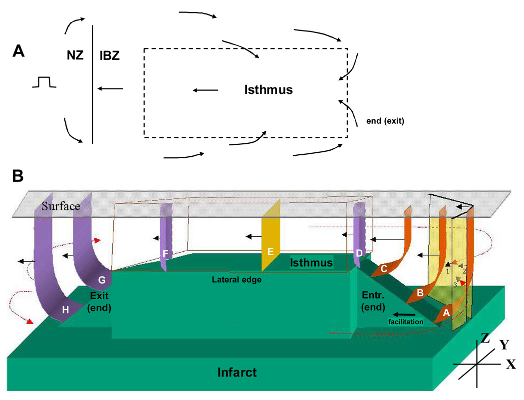
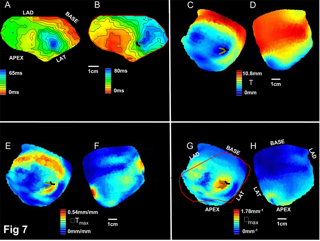
Methods: A geometric model describing the relationship between IBZ geometry and wavefront propagation in reentrant circuits was developed. Based on the formulation, slow conduction and block were expected to coincide with areas where IBZ thickness (T) is minimal and the local spatial gradient in thickness (DeltaT) is maximal, so that the degree of wavefront curvature rho proportional, variant DeltaT/T is maximal. Regions of fastest conduction velocity were predicted to coincide with areas of minimum DeltaT. In seven arrhythmogenic postinfarction canine heart experiments, tachycardia was induced by programmed stimulation, and activation maps were constructed from multichannel recordings. IBZ thickness was measured in excised hearts from histologic analysis or magnetic resonance imaging. Reentrant circuit properties were predicted from IBZ geometry and compared with ventricular activation maps after tachycardia induction.
Results: Mean IBZ thickness was 231 +/- 140 microm at the reentry isthmus and 1440 +/- 770 microm in the outer pathway (P <0.001). Mean curvature rho was 1.63 +/- 0.45 mm(-1) at functional block line locations, 0.71 +/- 0.18 mm(-1) at isthmus entrance-exit points, and 0.33 +/- 0.13 mm(-1) in the outer reentrant circuit pathway. The mean conduction velocity about the circuit during reentrant tachycardia was 0.32 +/- 0.04 mm/ms at entrance-exit points, 0.42 +/- 0.13 mm/ms for the entire outer pathway, and 0.64 +/- 0.16 mm/ms at outer pathway regions with minimum DeltaT. Model sensitivity and specificity to detect isthmus location was 75.0% and 97.2%.
Conclusions: Reentrant circuit features as determined by activation mapping can be predicted on the basis of IBZ geometrical relationships.
Figures


Comment in
-
Myths, metaphors, and mathematical models.Heart Rhythm. 2007 Aug;4(8):1046-7. doi: 10.1016/j.hrthm.2007.05.015. Epub 2007 May 21. Heart Rhythm. 2007. PMID: 17675079 No abstract available.
References
-
- Wit AL. Ablation of ventricular tachycardia: Does anyone have any new ideas? Heart Rhythm. 2006;3:198–200. - PubMed
-
- Garan H. A perspective on the ESVEM trial and current knowledge: Catheter ablation for ventricular tachyarrhythmias. Progress in Cardiovascular Diseases. 1996;38:457–462. - PubMed
-
- Wit AL, Allessie MA, Bonke FI, Lammers W, Smeets J, Fenoglio JJ., Jr Electrophysiologic mapping to determine the mechanism of experimental ventricular tachycardia initiated by premature impulses. Am J Cardiol. 1982;49:166–185. - PubMed
-
- Peters NS, Coromilas J, Severs NJ, Wit AL. Disturbed connexin43 gap junction distribution correlates with the location of reentrant circuits in the epicardial border zone of healing canine infarcts that cause ventricular tachycardia. Circulation. 1997;95:988–996. - PubMed
Publication types
MeSH terms
Grants and funding
LinkOut - more resources
Full Text Sources
Other Literature Sources
Medical
