. 2007 Aug;39(4):277-92.
doi: 10.1016/j.tice.2007.05.003. Epub 2007 Aug 6.

Organization, ultrastructure, and development of midgut visceral muscle in larval Aedes aegypti

Affiliations

Organization, ultrastructure, and development of midgut visceral muscle in larval Aedes aegypti

E P Bernick et al. Tissue Cell. 2007 Aug.

Abstract

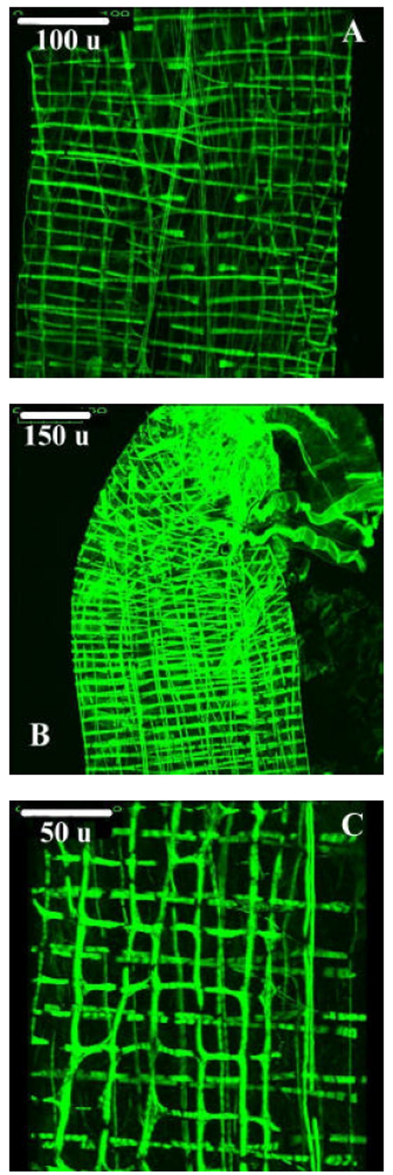
The midgut muscularis of larvae of the mosquito Aedes aegypti takes the form of a grid of longitudinal and circular muscle bands. The longitudinal and circular bands overlap at near right angles at many areas of intersection. The longitudinal bands run the length of the midgut. However, some bands of circular muscle, located in the anterior midgut, pass only partway around the gut. An unusual feature was observed at some regions where longitudinal and circular bands of muscle intersect: filaments oriented at near right angles to one another were present in the same membrane-bound fiber. These cruciform regions send contractile elements into both circular and longitudinal bands. The muscularis was fixed in a contracted state, so most of the sarcomeres are represented by complete overlap of myosin and lighter staining actin filaments. Features characteristic of supercontracting muscle, including perforated Z-lines, were seen in sarcomeres of circular muscle bands. Small invaginations resembling transverse tubules were present but a sarcoplasmic reticulum was not observed. While occasional cells that may be neurons or neurosecretory cells were observed, a network that might serve to coordinate the segmentation and peristaltic movement of the muscularis was not apparent.


Figures

FIGURE 1
FIGURE 1
Images of whole guts from the 2nd (A), 3rd (B) and 4th (C) instars of the mosquito Aedes aegypti. In each image, the anterior midgut is located in the upper right and the posterior midgut in the lower left. Each image was recorded at the same magnification (50x). Scale bars, located on the lower right margin of each image, represent 600 microns.
FIGURE 2
FIGURE 2
A: SEM micrograph of a 4th instar Aedes aegypti anterior midgut, magnification 700X. Regions of longitudinal muscle bands (L) overlapping circular bands (C) are present.Bifurcating circular bands of muscle (BC) are also present. Scale bar (lower right margin) represents 43 μm. B: SEM micrograph of a 2nd instar Aedes aegypti anterior midgut, magnification 500X. Cruciform regions of muscularis (A), containing both circular and longitudinal filaments within a single plane, are present in some specimens. Ovoid swellings (O) can be observed on both circular (C) and longitudinal (L) muscle bands. Gastric cecae (GC) are located along the upper border of the image. Scale bar (lower right margin) represents 60 μm.
FIGURE 3
FIGURE 3
A & B: SEM micrographs of branching circular muscle (BC) in the anterior (A) and posterior (B) midgut of a 3rd instar Aedes aegypti larva. Magnification in both micrographs is 400X. C= circular muscle; L= longitudinal muscle; GC= gastric cecae. Scale bars (lower right margin) represent 75 μm. C & D: SEM micrographs illustrating branching longitudinal muscle (BL) in the posterior midgut of a 4th instar Aedes aegypti larva. C= circular muscle; L= longitudinal muscle; MT= Malpighian tubule; HG= hindgut; PMG= posterior midgut. Scale bars (lower right margins) represent 86 μm (C) and 38 μm (D).
FIGURE 4
FIGURE 4
A: Longitudinal muscle (LM) extending onto the foregut (FG) of a 3rd instar Aedes aegypti larva. GC= gastric ceca; AMG= anterior midgut. Scale bar (lower right margin) represents 60 μm. B: Longitudinal muscle (L) extending onto the hindgut (HG) of a 3rd instar Aedes aegypti larva. MT= Malpighian tubules; PMG= posterior midgut; BL= branching longitudinal muscle. Scale bar (lower right margin) represents 75 μm.
FIGURE 5
FIGURE 5
Incomplete bands of circular muscle (IC) associated with the anterior midgut of a 4th instar Aedes aegypti larva. L= longitudinal muscle, C= circular muscle. Scale bar (lower right margin) represents 50 μm.
FIGURE 6
FIGURE 6
A: Cross section through a longitudinal muscle from the posterior midgut of a 2nd instar Aedes aegypti larva. Scale bar represents 1 μm. B: Cross section through a longitudinal muscle from the anterior midgut of a 3rd instar Aedes aegypti larva. Scale bar represents 1 μm. LM= Longitudinal muscle; SM= Suspending membrane; T= Tracheolar cell; ME= Midgut epithelium; BM= Basement membrane.
FIGURE 7
FIGURE 7
A: Cross section, longitudinal muscle from the anterior midgut of a 2nd instar Aedes aegypti larva. Scale bar represents 1 μm. B: Cross section, longitudinal muscle from the posterior midgut of a 3rd instar Aedes aegypti larva. Scale bar represents 0.5 μm. C: Cross section, longitudinal muscle from the posterior midgut of a 4th instar Aedes aegypti larva. Scale bar represents 0.5 μm. L= Longitudinal muscle; AMF= Actin & myosin filaments; SM= Sarcoplasmic membrane; MI= Membrane invagination; BM= Basement membrane; ME= Midgut epithelium; M= Mitochondria.
FIGURE 8
FIGURE 8
A: Longitudinal section, circular muscle from the anterior midgut of a 2nd instar Aedes aegypti larva. Scale bar represents 1 μm. B: Longitudinal section, circular muscle from the anterior midgut of a 3rd instar Aedes aegypti larva. Scale bar represents 2 μm. C: Longitudinal section, circular muscle from the anterior midgut of a 4th instar Aedes aegypti larva. Scale bar represents 5 μm. D: Longitudinal section, circular muscle from the anterior midgut of a 3rd instar Aedes aegypti larva. Scale bar represents 1 μm. CM= Circular muscle; LM= Longitudinal muscle; S= Sarcoplasm; Z= Z-lines; I= I bands; M= Mitochondria; T= Tracheolar cell; ME= Midgut epithelium.
FIGURE 9
FIGURE 9
A: Cross section through a cruciform cell from the anterior midgut of a 2nd instar Aedes aegypti larva. Scale bar represents 0.5 μm. B: Cross section through a cruciform cell from the anterior midgut of a 3rd instar Aedes aegypti larva. Scale bar represents 1 μm. C: Cross section through a cruciform cell from the anterior midgut of a 4th instar Aedes aegypti larva. Scale bar represents 1 μm. D: Cross section through a cruciform cell from the anterior midgut of a 4th instar Aedes aegypti larva, representing the lower portion of the muscle in Figure 9C at a higher magnification. Scale bar represents 0.5 μm. BM= Basement membrane; ME= Membrane epithelium; LF: Longitudinally oriented filaments; CF= Circularly oriented filaments; S= Sarcoplasm; M= Mitochondria.
FIGURE 10
FIGURE 10
A & B: Cross section through longitudinal muscle from the posterior midgut of a third instar Aedes aegypti larva. Membrane invaginations (MI) of the sarcoplasmic membrane (SM) are present. Actin and myosin filaments (AMF) are distinct. Mitochondria (M) are present in some muscle fibers. The basement membrane (BM) separates the muscle fibers from the midgut epithelium (ME). Scale bars represent 0.2 microns.
FIGURE 11
FIGURE 11
A: Three dimentional projection image from the anterior midgut of a 4th instar Aedes aegypti larva prepared for the confocal microscope using phalloidin conjugated to Alexa-Fluor 488. Bifurcations of circular muscle bands, as well as cruciform regions, can be identified. Scale bar (upper left corner) represents 100 microns. B: Three dimensional projection image from the posterior midgut of a 4th instar Aedes aegypti larva prepared for the confocal microscope using phalloidin. Bifurcating circular and longitudinal muscle bands can be identified. Scale bar (upper left corner) represents 150microns. C: Three dimensional projection image from the anterior midgut of a 4th instar Aedes aegypti larva prepared for the confocal microscope using phalloidin. Sharing of filaments between otherwise distinct bands of muscle can be observed. Longitudinal muscle bands are in the vertical orientation. Scale bar (upper left corner) represents 50 microns.
FIGURE 12
FIGURE 12
Illustration of excitation traveling along a longitudinal muscle band (L) associated with cruciform regions of muscularis. Circular muscle (C), anterior, and posterior orientation is indicated.

References

    1. Clark TM, et al. Additional Morphological and Physiological Heterogeneity Within the Midgut of Larval Aedes aegypti Revealed By Histology, Electrophysiology, and Effects of Bacillus thuringiensis Endotoxin. Tissue and Cell. 2005;37:457–468. - PubMed
    1. Clark TM, Koch A, Moffett DF. The Electrical Properties of the Anterior Stomach of the Larval Mosquito Aedes aegypti. Journal of Experimental Biology. 2000;203:1093–1101. - PubMed
    1. Clements AN. The Biology of Mosquitoes. Volume One. Chapman and Hall; New York, NY: 1992.
    1. Copenhaver PF, Horgan AM, Combes S. An Identified Set of Visceral Muscle Bands is Essential for the Guidance of Migratory Neurons in the Enteric Nervous System of Manduca sexta. Developmental Biology. 1996;179:412–426. - PubMed
    1. Copenhaver PF. Origins, Migration, and Differentiation of Glial Cells in the Insect Enteric Nervous System From a Discrete Set of Glial Precursors. Development. 1993;117:59–74.

Publication types