

Energy transfer in reconstituted peridinin-chlorophyll-protein complexes: ensemble and single-molecule spectroscopy studies
- PMID: 17675350
- PMCID: PMC2025647
- DOI: 10.1529/biophysj.107.112094
Energy transfer in reconstituted peridinin-chlorophyll-protein complexes: ensemble and single-molecule spectroscopy studies
Abstract
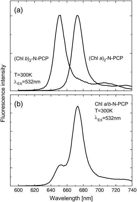
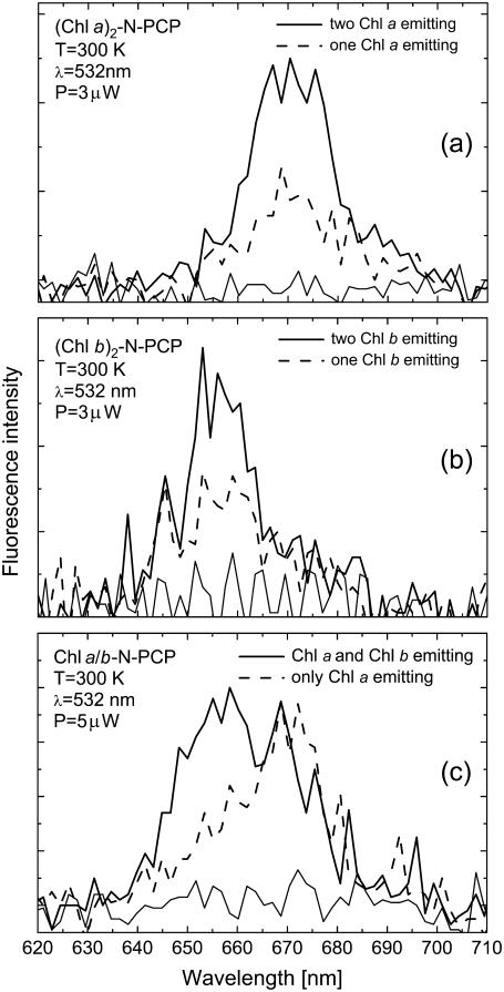
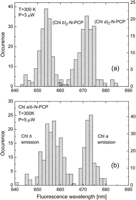
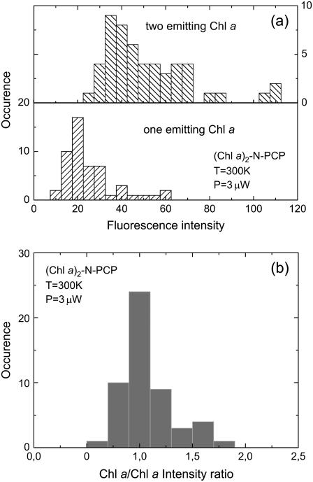
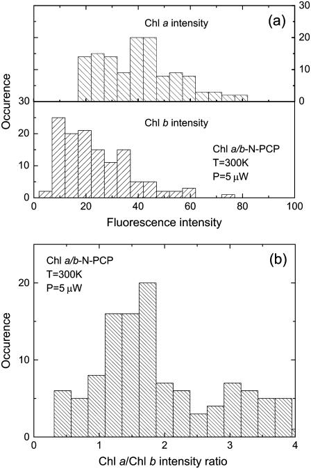
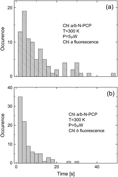
We combine ensemble and single-molecule spectroscopy to gain insight into the energy transfer between chlorophylls (Chls) in peridinin-chlorophyll-protein (PCP) complexes reconstituted with Chl a, Chl b, as well as both Chl a and Chl b. The main focus is the heterochlorophyllous system (Chl a/b-N-PCP), and reference information essential to interpret experimental observations is obtained from homochlorophyllous complexes. Energy transfer between Chls in Chl a/b-N-PCP takes place from Chl b to Chl a and also from Chl a to Chl b with comparable Förster energy transfer rates of 0.0324 and 0.0215 ps(-1), respectively. Monte Carlo simulations yield the ratio of 39:61 for the excitation distribution between Chl a and Chl b, which is larger than the equilibrium distribution of 34:66. An average Chl a/Chl b fluorescence intensity ratio of 66:34 is measured, however, for single Chl a/b-N-PCP complexes excited into the peridinin (Per) absorption. This difference is attributed to almost three times more efficient energy transfer from Per to Chl a than to Chl b. The results indicate also that due to bilateral energy transfer, the Chl system equilibrates only partially during the excited state lifetimes.
Figures

Similar articles
-
Monitoring fluorescence of individual chromophores in peridinin-chlorophyll-protein complex using single molecule spectroscopy.Biochim Biophys Acta. 2007 Jul;1767(7):956-64. doi: 10.1016/j.bbabio.2007.05.004. Epub 2007 May 18. Biochim Biophys Acta. 2007. PMID: 17572378
-
Tuning energy transfer in the peridinin-chlorophyll complex by reconstitution with different chlorophylls.Photosynth Res. 2005 Nov;86(1-2):217-27. doi: 10.1007/s11120-005-1447-x. Photosynth Res. 2005. PMID: 16172940
-
Carotenoid-to-chlorophyll energy transfer in recombinant major light-harvesting complex (LHCII) of higher plants. I. Femtosecond transient absorption measurements.Biophys J. 2001 Feb;80(2):901-15. doi: 10.1016/S0006-3495(01)76069-9. Biophys J. 2001. PMID: 11159457 Free PMC article.
-
Spectroscopy of the peridinin-chlorophyll-a protein: insight into light-harvesting strategy of marine algae.Arch Biochem Biophys. 2007 Feb 15;458(2):111-20. doi: 10.1016/j.abb.2006.10.006. Epub 2006 Oct 27. Arch Biochem Biophys. 2007. PMID: 17098207 Review.
-
Application of time-resolved polarization fluorescence spectroscopy in the femtosecond range to photosynthetic systems.Photochem Photobiol. 2007 Jan-Feb;83(1):163-70. doi: 10.1562/2006-02-28-IR-825. Photochem Photobiol. 2007. PMID: 16643087 Review.
Cited by
-
Light-induced energetic decoupling as a mechanism for phycobilisome-related energy dissipation in red algae: a single molecule study.PLoS One. 2008 Sep 4;3(9):e3134. doi: 10.1371/journal.pone.0003134. PLoS One. 2008. PMID: 18769542 Free PMC article.
-
Low-temperature spectral dynamics of single TDI molecules in n-alkane matrixes.J Fluoresc. 2008 May-Jul;18(3-4):625-31. doi: 10.1007/s10895-008-0326-1. Epub 2008 Feb 16. J Fluoresc. 2008. PMID: 18278543
-
Silver Island Film for Enhancing Light Harvesting in Natural Photosynthetic Proteins.Int J Mol Sci. 2020 Apr 1;21(7):2451. doi: 10.3390/ijms21072451. Int J Mol Sci. 2020. PMID: 32244795 Free PMC article. Review.
-
Fluorescence spectroscopy of reconstituted peridinin-chlorophyll-protein complexes.Photosynth Res. 2008 Feb-Mar;95(2-3):253-60. doi: 10.1007/s11120-007-9243-4. Epub 2007 Oct 31. Photosynth Res. 2008. PMID: 17972159
-
Energy transfer in the peridinin-chlorophyll protein complex reconstituted with mixed chlorophyll sites.Biophys J. 2008 Apr 15;94(8):3198-207. doi: 10.1529/biophysj.107.123430. Epub 2008 Jan 11. Biophys J. 2008. PMID: 18192358 Free PMC article.
References
-
- Tronrud, D. E., M. F. Schmid, and B. W. Matthews. 1986. Structure and x-ray amino-acid-sequence of a bacteriochlorophyll-a protein from prosthecochloris-aestuarii refined at 1.9 Å resolution. J. Mol. Biol. 188:443–454. - PubMed
-
- Zouni, A., H. T. Witt, J. Kern, P. Fromme, N. Krauss, W. Saenger, and P. Orth. 2001. Crystal structure of photosystem II from Synechococcus elongates at 3.8 angstrom resolution. Nature. 409:739–743. - PubMed
-
- Jordan, P., P. Fromme, H. T. Witt, O. Klukas, W. Saenger, and N. Krauss. 2001. Three-dimensional structure of cyanobacterial photosystem I at 2.5 angstrom resolution. Nature. 411:909–917. - PubMed
-
- Ben-Shem, A., F. Frolow, and N. Nelson. 2003. Crystal structure of plant photosystem I. Nature. 426:630–635. - PubMed
-
- Ferreira, K. N., T. M. Iverson, K. Maghlaoui, J. Barber, and S. Iwata. 2004. Architecture of the photosynthetic oxygen-evolving center. Science. 303:1831–1838. - PubMed
Publication types
MeSH terms
Substances
LinkOut - more resources
Full Text Sources
