

The archaeon Methanosarcina acetivorans contains a protein disulfide reductase with an iron-sulfur cluster
- PMID: 17675382
- PMCID: PMC2168450
- DOI: 10.1128/JB.00891-07
The archaeon Methanosarcina acetivorans contains a protein disulfide reductase with an iron-sulfur cluster
Abstract
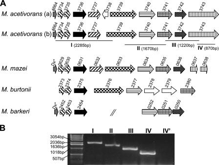
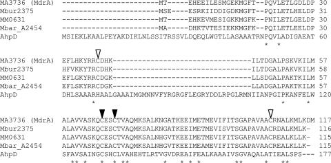
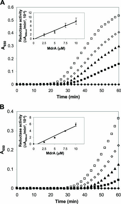
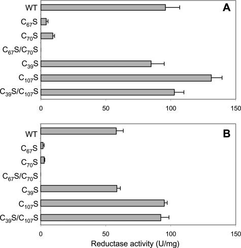
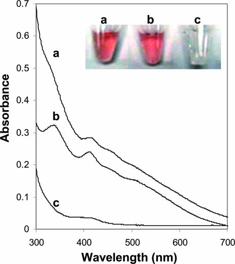
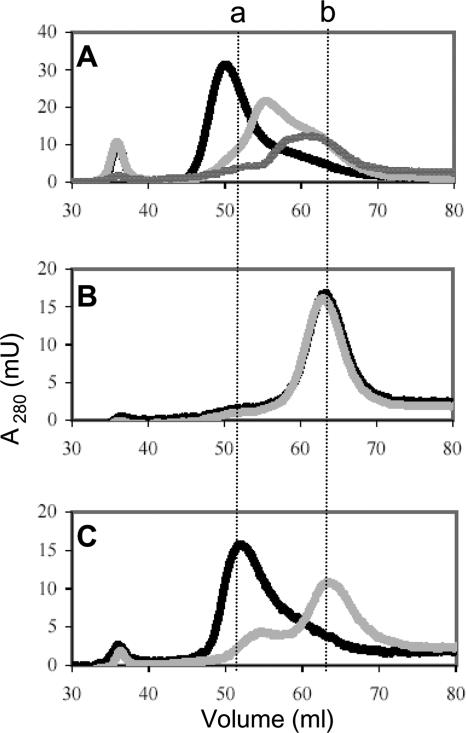
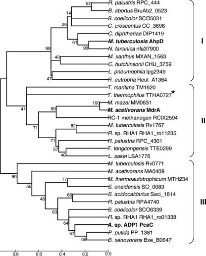
Methanosarcina acetivorans, a strictly anaerobic methane-producing species belonging to the domain Archaea, contains a gene cluster annotated with homologs encoding oxidative stress proteins. One of the genes (MA3736) is annotated as a gene encoding an uncharacterized carboxymuconolactone decarboxylase, an enzyme required for aerobic growth with aromatic compounds by species in the domain Bacteria. Methane-producing species are not known to utilize aromatic compounds, suggesting that MA3736 is incorrectly annotated. The product of MA3736, overproduced in Escherichia coli, had protein disulfide reductase activity dependent on a C(67)XXC(70) motif not found in carboxymuconolactone decarboxylase. We propose that MA3736 be renamed mdrA (methanosarcina disulfide reductase). Further, unlike carboxymuconolactone decarboxylase, MdrA contained an Fe-S cluster. Binding of the Fe-S cluster was dependent on essential cysteines C(67) and C(70), while cysteines C(39) and C(107) were not required. Loss of the Fe-S cluster resulted in conversion of MdrA from an inactive hexamer to a trimer with protein disulfide reductase activity. The data suggest that MdrA is the prototype of a previously unrecognized protein disulfide reductase family which contains an intermolecular Fe-S cluster that controls oligomerization as a mechanism to regulate protein disulfide reductase activity.
Figures
Similar articles
-
Methanosarcina acetivorans contains a functional ISC system for iron-sulfur cluster biogenesis.BMC Microbiol. 2020 Oct 23;20(1):323. doi: 10.1186/s12866-020-02014-z. BMC Microbiol. 2020. PMID: 33096982 Free PMC article.
-
Discovery and characterization of the first archaeal dihydromethanopterin reductase, an iron-sulfur flavoprotein from Methanosarcina mazei.J Bacteriol. 2014 Jan;196(2):203-9. doi: 10.1128/JB.00457-13. Epub 2013 Aug 30. J Bacteriol. 2014. PMID: 23995635 Free PMC article.
-
Structural and Biochemical Characterization of a Ferredoxin:Thioredoxin Reductase-like Enzyme from Methanosarcina acetivorans.Biochemistry. 2015 May 19;54(19):3122-8. doi: 10.1021/acs.biochem.5b00137. Biochemistry. 2015. PMID: 25915695
-
Structural and Biochemical Characterizations of Methanoredoxin from Methanosarcina acetivorans, a Glutaredoxin-Like Enzyme with Coenzyme M-Dependent Protein Disulfide Reductase Activity.Biochemistry. 2016 Jan 19;55(2):313-21. doi: 10.1021/acs.biochem.5b00823. Epub 2015 Dec 30. Biochemistry. 2016. PMID: 26684934
-
The roles of glutaredoxins ligating Fe-S clusters: Sensing, transfer or repair functions?Biochim Biophys Acta. 2015 Jun;1853(6):1513-27. doi: 10.1016/j.bbamcr.2014.09.018. Epub 2014 Sep 28. Biochim Biophys Acta. 2015. PMID: 25264274 Review.
Cited by
-
Assessment of the oxidant tolerance of Methanosarcina acetivorans.FEMS Microbiol Lett. 2013 Jun;343(1):13-9. doi: 10.1111/1574-6968.12115. Epub 2013 Mar 15. FEMS Microbiol Lett. 2013. PMID: 23448147 Free PMC article.
-
Genetic basis for metabolism of methylated sulfur compounds in Methanosarcina species.J Bacteriol. 2015 Apr;197(8):1515-24. doi: 10.1128/JB.02605-14. Epub 2015 Feb 17. J Bacteriol. 2015. PMID: 25691524 Free PMC article.
-
Dihydrophenylalanine: a prephenate-derived Photorhabdus luminescens antibiotic and intermediate in dihydrostilbene biosynthesis.Chem Biol. 2011 Sep 23;18(9):1102-12. doi: 10.1016/j.chembiol.2011.07.009. Chem Biol. 2011. PMID: 21944749 Free PMC article.
-
Genome-wide responses to carbonyl electrophiles in Bacillus subtilis: control of the thiol-dependent formaldehyde dehydrogenase AdhA and cysteine proteinase YraA by the MerR-family regulator YraB (AdhR).Mol Microbiol. 2009 Feb;71(4):876-94. doi: 10.1111/j.1365-2958.2008.06568.x. Epub 2008 Dec 23. Mol Microbiol. 2009. PMID: 19170879 Free PMC article.
-
Redox-sensitive DNA binding by homodimeric Methanosarcina acetivorans MsvR is modulated by cysteine residues.BMC Microbiol. 2013 Jul 16;13:163. doi: 10.1186/1471-2180-13-163. BMC Microbiol. 2013. PMID: 23865844 Free PMC article.
References
-
- Achebach, S., Q. H. Tran, A. Vlamis-Gardikas, M. Mullner, A. Holmgren, and G. Unden. 2004. Stimulation of Fe-S cluster insertion into apoFNR by Escherichia coli glutaredoxins 1, 2 and 3 in vitro. FEBS Lett. 565:203-206. - PubMed
-
- Alam, M. S., S. K. Garg, and P. Agrawal. 2007. Molecular function of WhiB4/Rv3681c of Mycobacterium tuberculosis H37Rv: a [4Fe-4S] cluster co-ordinating protein disulphide reductase. Mol. Microbiol. 63:1414-1431. - PubMed
-
- Auchere, F., S. R. Pauleta, P. Tavares, I. Moura, and J. J. Moura. 2006. Kinetics studies of the superoxide-mediated electron transfer reactions between rubredoxin-type proteins and superoxide reductases. J. Biol. Inorg. Chem. 11:433-444. - PubMed
-
- Beinert, H. 1983. Semi-micro methods for analysis of labile sulfide and of labile sulfide plus sulfane sulfur in unusually stable iron-sulfur proteins. Anal. Biochem. 131:373-378. - PubMed
-
- Berndt, C., C. Hudemann, E. M. Hanschmann, R. Axelsson, A. Holmgren, and C. H. Lillig. 2007. How does iron-sulfur cluster coordination regulate the activity of human glutaredoxin 2? Antioxid. Redox Signal. 9:151-157. - PubMed
Publication types
MeSH terms
Substances
Grants and funding
LinkOut - more resources
Full Text Sources
Other Literature Sources
Molecular Biology Databases
Miscellaneous
