. 2007 Nov;189(22):7961-7.
doi: 10.1128/JB.00789-07. Epub 2007 Aug 3.

Generation of virulence factor variants in Staphylococcus aureus biofilms

Affiliations

Generation of virulence factor variants in Staphylococcus aureus biofilms

Jeremy M Yarwood et al. J Bacteriol. 2007 Nov.

Abstract

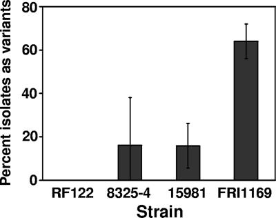
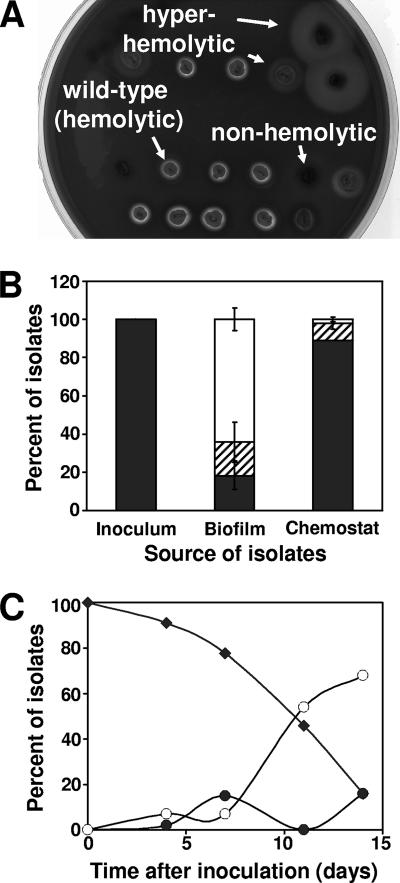
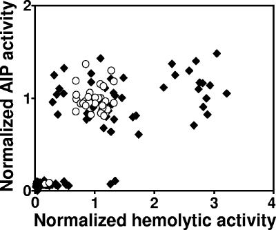
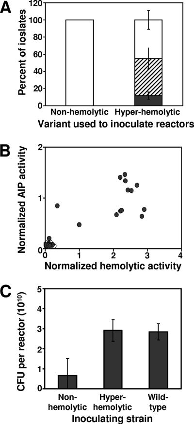
Several serious diseases are caused by biofilm-associated Staphylococcus aureus. Colonial variants occur in biofilms of other bacterial species, and S. aureus variants are frequently isolated from biofilm-associated infections. Thus, we studied the generation of variants with altered expression of virulence factors in S. aureus biofilms. We observed that the number of variants found in biofilms, as measured by hemolytic activity, varied for different strains. Further study of hemolytic activity and signaling by the accessory gene regulator (Agr) quorum-sensing system in one S. aureus strain revealed three primary biofilm subpopulations: nonhemolytic (Agr deficient), hemolytic (Agr positive), and hyperhemolytic (also Agr positive). The nonhemolytic variant became the numerically dominant subpopulation in the biofilm. The nonhemolytic variant phenotype was stable and heritable, indicating a genetic perturbation, whereas the hyperhemolytic phenotype was unstable, suggesting a phase variation. Transcription profiling revealed that expression of the agr locus and many extracellular virulence factors was repressed in the nonhemolytic variant. Expression of the agr-activating gene, sarU, was also repressed in the nonhemolytic variant, suggesting one potential regulatory pathway responsible for the Agr-deficient phenotype. We suggest that the development of these variants in biofilms may have important clinical implications.


Figures

FIG. 1.
FIG. 1.
Hemolytic variant formation in isolates of various S. aureus strains grown in drip reactor biofilms for 2 weeks as determined by plating on sheep blood agar. Values represent averages and standard deviations of variant percentages in four biofilm cultures of each strain. The value for FRI1169 is significantly higher than for the other strains (P < 0.01, Student t test).
FIG. 2.
FIG. 2.
Hemolytic variant generation in cultures of S. aureus FRI1169. (A) Isolates from drip reactors plated onto sheep blood agar. (B) Distribution of hemolytic (▪), hyperhemolytic (▒), and nonhemolytic (□) isolates in overnight culture used to inoculate drip reactors, 2-week-old drip reactor biofilms, and 2-week-old chemostat (planktonic) cultures. Values represent averages and standard deviations of three independent cultures. Changes in the hemolytic and nonhemolytic (but not the hyperhemolytic) populations between the biofilm and chemostat cultures are significant (P < 0.001, Student t test). (C) Hemolytic (⧫), hyperhemolytic (•), and nonhemolytic (○) isolates from drip reactor biofilms sampled at 0, 4, 11, and 14 days after inoculation.
FIG. 3.
FIG. 3.
Hemolytic and AIP-signaling activity of isolates from biofilm (⧫) and chemostat (○) cultures. Values are normalized to activities measured in wild-type FRI1169.
FIG. 4.
FIG. 4.
Variant generation in 2-week biofilms inoculated with nonhemolytic and hyperhemolytic variants. (A) Distribution of hemolytic (▪), hyperhemolytic (▒), and nonhemolytic (□) isolates from drip reactors inoculated with indicated variant as determined by plating on sheep blood agar. Values represent averages and standard deviations of variant percentages in four biofilm cultures inoculated with each variant. (B) Hemolytic and AIP-signaling activity of isolates from biofilms inoculated with nonhemolytic (○) or hyperhemolytic (•) variants. (C) Viable cells recovered from reactors inoculated with indicated variant. Values represent averages and standard deviations of variant percentages in four biofilm cultures inoculated with each variant. Differences between the values for the hyperhemolytic or wild-type strain and the nonhemolytic strain are significant (P < 0.01, Student t test), while there is no significant difference between the values for the hyperhemolytic and wild-type strains (P = 0.87, Student t test).

Similar articles

Cited by

References

    1. Akiyama, H., T. Hamada, W. K. Huh, O. Yamasaki, T. Oono, W. Fujimoto, and K. Iwatsuki. 2003. Confocal laser scanning microscopic observation of glycocalyx production by Staphylococcus aureus in skin lesions of bullous impetigo, atopic dermatitis, and pemphigus foliaceus. Br. J. Dermatol. 148:526-532. - PubMed
    1. Allegrucci, M., and K. Sauer. 2007. Characterization of colony morphology variants isolated from Streptococcus pneumoniae biofilms. J. Bacteriol. 189:2030-2038. - PMC - PubMed
    1. Bergdoll, M. S., B. A. Crass, R. F. Reiser, R. N. Robbins, and J. P. Davis. 1981. A new staphylococcal enterotoxin, enterotoxin F, associated with toxic-shock-syndrome Staphylococcus aureus isolates. Lancet 1:1017-1021. - PubMed
    1. Bernheimer, A. W. 1988. Assay of hemolytic toxins. Methods Enzymol. 165:213-217. - PubMed
    1. Boles, B. R., M. Thoendel, and P. K. Singh. 2004. Self-generated diversity produces “insurance effects” in biofilm communities. Proc. Natl. Acad. Sci. USA 101:16630-16635. - PMC - PubMed

Publication types

MeSH terms

Associated data