. 2007 Sep;73(18):5797-808.
doi: 10.1128/AEM.00146-07. Epub 2007 Aug 3.

Characterization of protein-protein interactions involved in iron reduction by Shewanella oneidensis MR-1

Affiliations

Characterization of protein-protein interactions involved in iron reduction by Shewanella oneidensis MR-1

Daniel E Ross et al. Appl Environ Microbiol. 2007 Sep.

Abstract

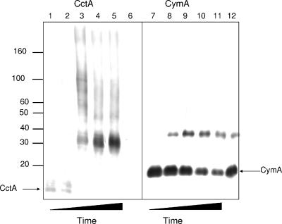
The interaction of proteins implicated in dissimilatory metal reduction by Shewanella oneidensis MR-1 (outer membrane [OM] proteins OmcA, MtrB, and MtrC; OM-associated protein MtrA; periplasmic protein CctA; and cytoplasmic membrane protein CymA) were characterized by protein purification, analytical ultracentrifugation, and cross-linking methods. Five of these proteins are heme proteins, OmcA (83 kDa), MtrC (75 kDa), MtrA (32 kDa), CctA (19 kDa), and CymA (21 kDa), and can be visualized after sodium dodecyl sulfate-polyacrylamide gel electrophoresis by heme staining. We show for the first time that MtrC, MtrA, and MtrB form a 198-kDa complex with a 1:1:1 stoichiometry. These proteins copurify through anion-exchange chromatography, and the purified complex has the ability to reduce multiple forms of Fe(III) and Mn(IV). Additionally, MtrA fractionates with the OM through sucrose density gradient ultracentrifugation, and MtrA comigrates with MtrB in native gels. Protein cross-linking of whole cells with 1% formaldehyde show new heme bands of 160, 151, 136, and 59 kDa. Using antibodies to detect each protein separately, heme proteins OmcA and MtrC were shown to cross-link, yielding the 160-kDa band. Consistent with copurification results, MtrB cross-links with MtrA, forming high-molecular-mass bands of approximately 151 and 136 kDa.


Figures

FIG. 1.
FIG. 1.
Western blot of whole-cell extracts from S. oneidensis MR-1 using affinity-purified antibodies to CctA. Whole cells were sonicated, and 8-μg samples of proteins was loaded onto 12% SDS-polyacrylamide gels. The positions of molecular mass markers (in kilodaltons) are indicated to the left of the gel.
FIG. 2.
FIG. 2.
(A) Q-Sepharose anion-exchange chromatography profile of TM fractions from S. oneidensis MR-1 monitored at heme-associated absorbance at 410 nm. (B) Western blots of protein fractions from Q-Sepharose column indicating the elution profiles of OmcA, MtrC, MtrB, and MtrA. Four replicate gels containing 12-μl amounts of protein from fractions 26 to 60 (even-numbered fractions correspond to those labeled on the elution profile) were run on SDS-polyacrylamide gels and transferred to nitrocellulose membranes. Detection of antibodies used Immobilon Western AP substrate. Note that fraction 50 for the MtrC visualization is lower in intensity due to leakage of the well.
FIG. 3.
FIG. 3.
Gel permeation chromatography of the MtrC/A/B peak fractions and analysis by SDS-PAGE. Fractions 38 to 56 shown in Fig. 2 were pooled, concentrated, and applied to a gel filtration column. (A) Elution profile of MtrC/A/B from gel filtration (Sephacryl S-300 HR resin; Amersham Biosciences) as monitored from the heme absorbance at 409 nm. The arrow indicates the void volume (blue dextran). (B) 10% SDS-PAGE of MtrC/A/B fraction taken from the peak fraction after gel filtration and visualized with either silver staining (lane 1) or heme staining (lane 2). The predominant heme peaks correspond to MtrC (69 kDa) and MtrA (30 kDa). The positions of molecular mass markers (in kilodaltons) are indicated to the left of the gel.
FIG. 4.
FIG. 4.
(A) Western blots of MtrC/A/B proteins separated by native PAGE. Fractions 38 to 56 shown in Fig. 2 containing the peak MtrC/A/B fractions were pooled, and 15-μl samples were applied to the gel. OM proteins MtrC, MtrB, and MtrA were probed with polyclonal antibodies. (B) TM proteins (15 μg per lane) separated using native PAGE and probed for OmcA and MtrC. Protein bands are indicated with arrows at the sides of the gels.
FIG. 5.
FIG. 5.
(A) Analytical ultracentrifugation (sedimentation equilibrium) analysis of MtrC/A/B. A representative plot of absorbance profiles (measured at an absorbance value of 530 nm) of 2.5 μM MtrC/A/B following centrifugation runs at 6,000 (squares), 8,000 (triangles), and 10,000 rpm (diamonds) (16 h at 20°C). The data from all three centrifugation speeds were simultaneously fitted to the equation for a single-species, noninteracting model (solid lines). Conditions of measurement were MtrC/A/B samples in 50 mM HEPES, 200 mM NaCl, 5% (wt/vol) Triton X-100, pH 7.6. Residuals between the experimental data and the fitted lines are shown in the graph at the top of the figure. (B) Representative visualization of sedimentation equilibrium profiles of two concentrations of MtrC/A/B, 2.5 μM (squares) and 0.25 μM (circles) measured at absorbance values of 530 nm and 440 nm, respectively, following centrifugation at 6000 rpm for 16 h at 20°C. Plots of ln absorbance versus (r2r02)/2 of both MtrC/A/B samples display similar straight-line slopes, demonstrating single-species, homogeneous system and a derived molecular mass that is independent of concentration in the range examined. Conditions of measurement were MtrC/A/B samples in 50 mM HEPES, 200 mM NaCl, 5% (wt/vol) Triton X-100, pH 7.6. Residuals between the experimental data and the fitted lines are shown in the graph at the top of the figure.
FIG. 6.
FIG. 6.
Reduction of various iron and manganese forms with chemically reduced MtrC/A/B. MtrC/A/B from the Sephacryl S-300 column was reduced by dithionite. To the reduced complex, we added either no iron (control), ferric citrate (1 mM), goethite (1 mg/ml), ferrihydrite (2 mM), or birnessite (1 mg/ml). The no-iron addition was scanned at 0, 10, 20, 30, and 40 min, whereas the other samples were scanned at the time intervals shown in the figures. The arrow indicates direction of spectral change going from fully reduced to fully oxidized.
FIG. 7.
FIG. 7.
(A) Separation of proteins from S. oneidensis MR-1 cultures into the CM, OM, and TM fractions. MtrA is shown to fractionate with the OM as does MtrB. CymA associates with the cytosolic membrane. (B) Heme staining of S. oneidensis cell extracts (lane 1), supernatant of washed cells (lane 2), and periplasmic fraction (lane 3). (C) Western blot using anti-MtrA of the same fractions as in panel B. The positions of molecular mass markers (in kilodaltons) are indicated to the left of the gel in panels B and C.
FIG. 8.
FIG. 8.
Heme staining of cells cross-linked by 1% formaldehyde and transferred to a nitrocellulose membrane. Lane 1, untreated cells that were boiled; lane 2, cells treated for 0 min; lane 3, cells treated for 30 min; lane 4, cells treated for 60 min; lane 5, cells treated for 120 min; lane 6, cells treated for 120 min and boiled. Each lane was loaded with 1 absorbance unit of cells to increase the concentration of low-abundance heme proteins. The arrows on the left indicate the bands that are observed, and the heme proteins OmcA, MtrC, MtrA, and CymA are identified. The new heme protein bands as a result of treatment with formaldehyde are indicated by arrows without labels. The positions of molecular mass markers (in kilodaltons) are indicated to the right of the membrane.
FIG. 9.
FIG. 9.
In vivo cross-linking of cells grown with ferric citrate as the terminal electron acceptor probed for complexes containing OmcA (A), MtrC (B), MtrB (C), MtrA (D), and CymA (E). Lanes 1, untreated cells that were boiled; lanes 2 to 5, cells treated with 1% formaldehyde for 0 min (lanes 2), 30 min (lanes 3), 60 min (lanes 4), and 120 min (lanes 5); lanes 6, cells treated with 1% formaldehyde for 120 min and boiled. The positions of formaldehyde cross-linked complexes are indicated by arrows to the left of the gels, and the molecular mass markers are indicated to the right of the gels.
FIG. 10.
FIG. 10.
In vivo cross-linking of cells grown with ferric citrate as the terminal electron acceptor probed for complexes containing CctA and CymA. Lanes 1 and 7, untreated cells and boiled; lanes 2 and 8, cells treated with 1% formaldehyde for 0 min; lanes 3 and 9, cells treated with 1% formaldehyde for 30 min; lanes 4 and 10, cells treated with 1% formaldehyde for 60 min; lanes 5 and 11, cells treated with 1% formaldehyde for 120 min; lanes 6 and 12, cells treated with 1% formaldehyde for 120 min and boiled. The positions of formaldehyde cross-linked complexes are indicated by arrows at the sides of the gels, and the positions of molecular mass markers (in kilodaltons) are indicated to the left of the gel.
FIG. 11.
FIG. 11.
Model depicting the interactions of OM proteins as a result of cross-linking and native protein methods. Interactions of CymA and CctA are yet to be defined. Menaquinone is shown in the membrane and shown as “Q” in the figure. The interaction of MtrC/A/B as a complex has been demonstrated in this work. We and others (52) have demonstrated the interaction of OmcA and MtrC. MtrA faces the periplasm, while OmcA and MtrC are extracellularly exposed lipoproteins and may potentially contact mineral oxides. The reduction potentials of various proteins and electron acceptors are shown to the left. Reduction potentials were obtained from the following: formate (62), menaquinone (42), MtrA (44), CctA (11), EDTA-Fe3+ (49), NTA-Fe3+, goethite, and citrate-Fe3+ (58), and pyrolusite (55). Decoupling of the electron transfer to insoluble TEAs from the proton pump at the CM implies that electron transfer to solid oxides does not provide energy to the cell but serves only to ground the cell by dumping electrons.

References

    1. Arnold, R. G., T. J. DiChristina, and M. R. Hoffmann. 1986. Inhibitor studies of dissimilative Fe(III) reduction by Pseudomonas sp. strain 200 (“Pseudomonas ferrireductans”). Appl. Environ. Microbiol. 52:281-289. - PMC - PubMed
    1. Arslan, E., H. Schulz, R. Zufferey, P. Kunzler, and L. Thony-Meyer. 1998. Overproduction of the Bradyrhizobium japonicum c-type cytochrome subunits of the cbb3 oxidase in Escherichia coli. Biochem. Biophys. Res. Commun. 251:744-747. - PubMed
    1. Beliaev, A. S., D. M. Klingeman, J. A. Klappenbach, L. Wu, M. F. Romine, J. M. Tiedje, K. H. Nealson, J. K. Fredrickson, and J. Zhou. 2005. Global transcriptome analysis of Shewanella oneidensis MR-1 exposed to different terminal electron acceptors. J. Bacteriol. 187:7138-7145. - PMC - PubMed
    1. Beliaev, A. S., and D. A. Saffarini. 1998. Shewanella putrefaciens mtrB encodes an outer membrane protein required for Fe(III) and Mn(IV) reduction. J. Bacteriol. 180:6292-6297. - PMC - PubMed
    1. Beliaev, A. S., D. A. Saffarini, J. L. McLaughlin, and D. Hunnicutt. 2001. MtrC, an outer membrane decahaem c cytochrome required for metal reduction in Shewanella putrefaciens MR-1. Mol. Microbiol. 39:722-730. - PubMed

Publication types

LinkOut - more resources