. 2007 Sep;171(3):838-45.
doi: 10.2353/ajpath.2007.070008. Epub 2007 Aug 3.

gamma delta T cells are necessary for platelet and neutrophil accumulation in limbal vessels and efficient epithelial repair after corneal abrasion

Affiliations

gamma delta T cells are necessary for platelet and neutrophil accumulation in limbal vessels and efficient epithelial repair after corneal abrasion

Zhijie Li et al. Am J Pathol. 2007 Sep.

Abstract

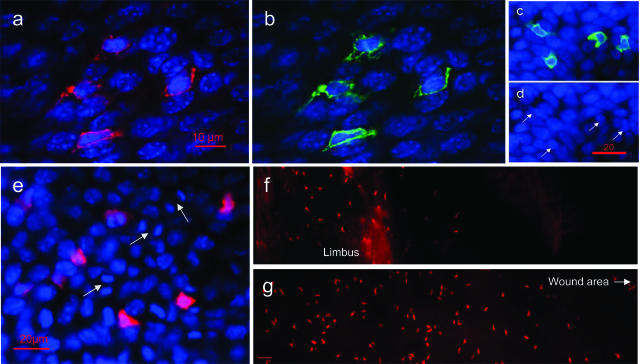
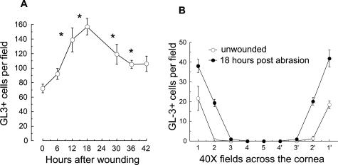
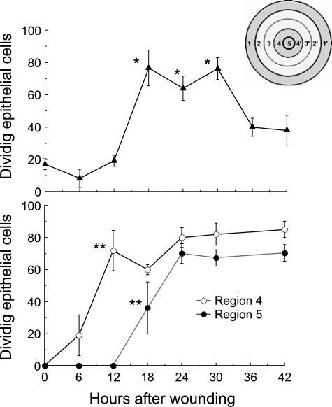
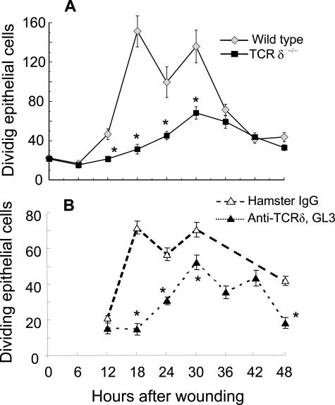
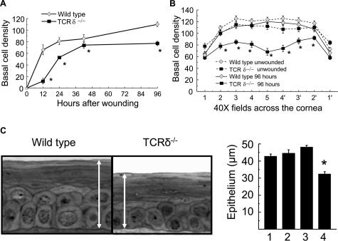
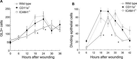
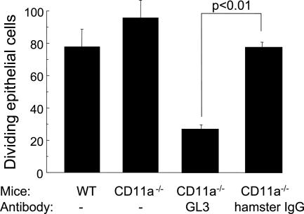
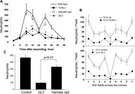
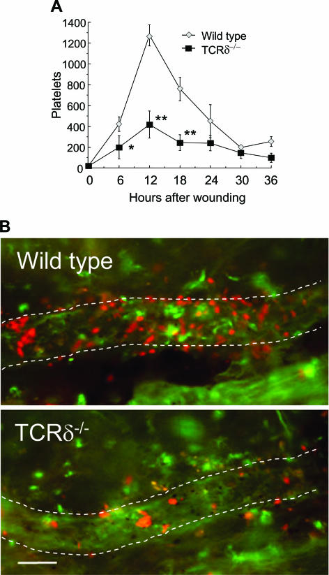
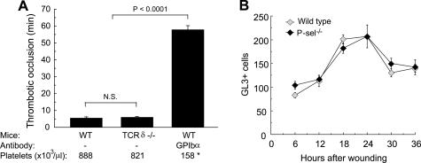
Corneal epithelial abrasion in C57BL/6 mice induces an inflammatory response with peak accumulation of neutrophils in the corneal stroma within 12 hours. Platelets localize in the limbal vessels throughout the same time course as neutrophils and contribute to wound healing because antibody-dependent depletion of platelets retards epithelial division and wound closure. In the present study, T cells in the limbal epithelium were found to predominantly express the gammadelta T-cell receptor (TCR). Corneal abrasion in wild-type, CD11a(-/-), and P-sel(-/-) mice increased the numbers of gammadelta T cells in the limbal and peripheral corneal epithelium and in the corneal stroma adjacent to the limbal blood vessels. Intercellular adhesion molecule (ICAM)-1(-/-) mice exhibited a reduction in gammadelta T-cell accumulation. TCRdelta(-/-) mice exhibited reduced inflammation and delayed epithelial wound healing as evidenced by delayed wound closure, reduced epithelial cell division, reduced neutrophil infiltration, and reduced epithelial cell density at 96 hours after wounding. TCRdelta(-/-) mice also exhibited >60% reduction in platelet localization in the limbus despite similar platelet counts and platelet function assessed with an in vivo thrombosis model. These results are consistent with the conclusion that gammadelta T cells are necessary for efficient inflammation, platelet localization in the limbus, and epithelial wound healing after corneal abrasion.


Figures

Figure 1
Figure 1
Immunofluorescence studies of murine corneal T cells. a: Unwounded corneal epithelium from a wild-type C57BL/6 mouse stained with PE-labeled anti-TCRδ, GL3. b: FITC-labeled anti-CD3 showing four cells that are positive for both antibodies. Nuclei are stained with DAPI. Unwounded corneal epithelium from TCRδ−/− mice stained with FITC-labeled anti-CD3 (c) and PE-labeled anti-TCRδ, GL3 (d). The arrows indicate the positions of the four CD3+ cells demonstrating the absence of TCRδ expression. e: Wild-type C57BL/6 mouse cornea at 18 hours after central corneal abrasion. The field of view is within limbal epithelium (see region 1 of Figure 3) showing PE-labeled anti-TCRδ (GL3)-stained cells in the region of dividing cells (arrows). Nuclei and mitotic figures stained with DAPI. f: Unwounded limbal and peripheral corneal epithelium from a wild-type mouse stained with PE-labeled anti-TCRδ, GL3 showing the restricted distribution of the GL3+ cells to the limbus. g: Limbal and peripheral corneal epithelium 18 hours after central corneal abrasion stained with PE-labeled anti-TCRδ (GL3) showing an increased number of GL3+ cells and expanded distribution of the GL3+ cells into the cornea. Scale bars: 10 μm (a and b); 20 μm (c and d); 40 μm (f and g).
Figure 2
Figure 2
Changes in GL3+ cells after central corneal epithelial abrasion. A: Temporal changes in the number of GL3+ cells after corneal abrasion in C57BL/6 wild-type mice. The values plotted at each time point are the sum of GL3+ cells in the epithelium and stroma in nine ×40 fields of view across the diameter of the cornea (n = 4). *P < 0.01 compared with unwounded corneas. B: The distribution of GL3+ cells at 18 hours after abrasion is plotted as cells per ×40 fields across the diameter of the cornea (limbus to limbus).
Figure 3
Figure 3
Epithelial response to central corneal epithelial abrasion in C57BL/6 mice—morphometric analysis. Inset: Schematic representation of whole mount excised cornea showing regions for microscopic analysis from the limbus (region 1) to the original wound area (regions 4 and 5). The width of each region is encompassed by the field of view of ×40 objective (∼0.53 mm). Basal epithelial cell division is the total number of dividing cells counted in three ×40 fields (regions 1, 2, and 3) across the cornea in the epithelium not directly injured during the central abrasion. Basal epithelial cell density is the number of cells per ×40 field in region 4 (the peripheral region abraded) and in region 5 (the center of the abraded area). *P < 0.01 compared with the level of cell division in uninjured corneas; **P < 0.01 compared with the density immediately after abrasion, n = 6.
Figure 4
Figure 4
Corneal response to central corneal epithelial abrasion in TCRδ−/− mice. Dividing basal epithelial cells were evaluated at 6-hour intervals after central corneal abrasion in wild-type mice and TCRδ−/− mice (A), and wild-type mice treated before wounding with either GL3 antibody or hamster IgG (B). The values for the dividing epithelial cells are sums of six ×40 fields of view (regions 1, 2, and 3 shown in the schema in Figure 3). The lower number of dividing cells in controls on B compared with the wild type in A reflects mouse strain differences. These antibody-blocking studies were done with C57BL/6 mice from Harlan (Indianapolis, IN). The studies in A were with C57BL/6 mice from The Jackson Laboratory, the appropriate controls for the TCRδ−/− mice. Statistical analysis indicated the values for the TCRδ−/− mice (n = 4) at 12, 18, 24, and 30 hours after wounding were significantly less than wild type (P < 0.01), and that for animals (n = 4) treated with GL3 (anti-TCRδ), values at 18, 24, 30, and 48 hours after wounding were significantly less than (P < 0.01) the hamster IgG controls.
Figure 5
Figure 5
Corneal epithelial healing after central abrasion in wild-type and TCRδ−/− mice. A: Basal epithelial density was determined in region 4 shown in the schema in Figure 3, ie, the periphery of the epithelial wound. *P < 0.01, n = 4. B: Basal cell density was determined at 96 hours after central abrasion. Values from ×40 fields across the diameter of the cornea are shown. *P < 0.01. C: Analysis of epithelial thickness in the center of the cornea at 96 hours after abrasion shown in photomicrographs of representative wild-type and TCRδ−/− epithelium. Plots of epithelial thickness in region 5 measured from toluidine blue-stained plastic sections of wild-type corneas (1), unwounded; wild-type corneas at 96 hours after abrasion (2); TCRδ−/− corneas, unwounded (3); and TCRδ−/− corneas at 96 hours after abrasion (4), *P < 0.01, n = 4.
Figure 6
Figure 6
Response to corneal abrasion in knockout mice. A: Mice with targeted deletions of CD11a or ICAM-1 were evaluated for GL3+ cell accumulation (expressed as sums of cells in nine ×40 fields of view across a diameter of the cornea) after central corneal epithelial abrasion. CD11a−/− mice were very similar to wild-type mice in this parameter. ICAM-1−/− mice exhibited significant reductions in GL3+ cell accumulation at 18 and 24 hours after wounding when compared with wild-type mice (*P < 0.001, n = 4). B: Mice with targeted deletions of CD11a or ICAM-1 were evaluated for epithelial cell division (sums of three ×40 fields of view, regions 1, 2, and 3 shown in the schema in Figure 3) after central corneal epithelial abrasion. CD11a−/− mice were similar to wild-type mice in this parameter. ICAM-1−/− mice exhibited significant reductions in epithelial cell division at 18, 30, and 36 hours after wounding (*P < 0.001, n = 4).
Figure 7
Figure 7
Effect of GL3 on epithelial division in CD11a−/− mice. CD11a−/− mice were treated with antibody GL3 or hamster IgG before central corneal epithelial abrasion. At 18 hours after wounding, corneas were collected and evaluated for epithelial cell division (n = 4).
Figure 8
Figure 8
Leukocyte emigration after central corneal abrasion. A: Neutrophil accumulation kinetics throughout a 48-hour observation time based on analysis of corneas collected at 6-hour intervals from wild-type mice, TCRδ−/− mice, and wild-type mice treated before wounding with either GL3 antibody or hamster IgG. Plotted is the sum of the number of neutrophils in nine fields of view across a diameter of the cornea, see Figure 3. *P < 0.01 for both TCRδ−/− mice and wild-type mice treated with GL3 compared with appropriate controls at 12, 18, 30, 36, and 42 hours after wounding, n = 4. B: Neutrophil (per nine fields of view) distributions across the corneas collected at 12 hours after wounding. All values for TCRδ−/− mice and wild-type mice treated with GL3 are significantly lower (P < 0.01, n = 4) compared with appropriate controls, except in region 5. C: Wild-type mice were given antibody GL3 at 18 hours after central corneal epithelial abrasion. Corneas were collected at 30 hours after abrasion and evaluated for neutrophil emigration (expressed as the sum of neutrophils counted in nine fields of view across a diameter of the cornea, n = 4).
Figure 9
Figure 9
Platelet accumulation in TCRδ−/− mice after corneal abrasion. A: Corneas were evaluated for platelet accumulation (expressed as the sum of platelets in eight fields of view in the limbal region) at 6-hour intervals after central corneal epithelial abrasion (*P < 0.05, **P < 0.01; n = 4). B: Photomicrographs of the limbus showing wild-type and TCRδ−/− mice 12 hours after epithelial abrasion stained with FITC-labeled anti-CD31 (labels endothelial junctions and some neutrophils) and PE-labeled anti-CD41 (to identify platelets). Scale bar, 25 μm.
Figure 10
Figure 10
A: Evaluation of platelet function in TCRδ−/− mice. Light dye-induced thrombotic venous occlusion was induced in wild-type (WT) and TCRδ−/− mice. The time from vessel injury until blood flow stopped is plotted. As a control setting, WT mice received anti-GP1bα to induce thrombocytopenia, as shown by the platelet counts (asterisk). n = 4. B: Accumulation of GL3+ cells in P-selectin-deficient mice. Because P-selectin-deficient (P-sel−/−) mice exhibit marked reductions in platelet and neutrophil accumulation in the limbal vessels after corneal abrasion, the effects of P-selectin deficiency on GL3+ cell accumulation in the epithelium and stroma (expressed as sums of cells in nine ×40 fields of view across a diameter of the cornea) were evaluated at 6-hour intervals. Statistical analysis revealed no difference in these curves, n = 4.

Similar articles

Cited by

References

    1. O’Brien TP, Li Q, Ashraf MF, Matteson DM, Stark WJ, Chan CC. Inflammatory response in the early stages of wound healing after excimer laser keratectomy. Arch Ophthalmol. 1998;116:1470–1474. - PubMed
    1. Wilson SE, Mohan RR, Mohan RR, Ambrosio R, Jr, Hong J, Lee J. The corneal wound healing response: cytokine-mediated interaction of the epithelium, stroma, and inflammatory cells. Prog Retin Eye Res. 2001;20:625–637. - PubMed
    1. Zhu SN, Dana MR. Expression of cell adhesion molecules on limbal and neovascular endothelium in corneal inflammatory neovascularization. Invest Ophthalmol Vis Sci. 1999;40:1427–1434. - PubMed
    1. Zhao M, Song B, Pu J, Forrester JV, McCaig CD. Direct visualization of a stratified epithelium reveals that wounds heal by unified sliding of cell sheets. FASEB J. 2003;17:397–406. - PMC - PubMed
    1. Savill J. Apoptosis in resolution of inflammation. J Leukoc Biol. 1997;61:375–380. - PubMed

Publication types

MeSH terms

Substances