Review
. 2007 Aug 28;46(34):9665-73.
doi: 10.1021/bi7010064. Epub 2007 Aug 3.

Interactions that drive Sec-dependent bacterial protein transport

Affiliations
Review

Interactions that drive Sec-dependent bacterial protein transport

Sharyn L Rusch et al. Biochemistry. .

Abstract

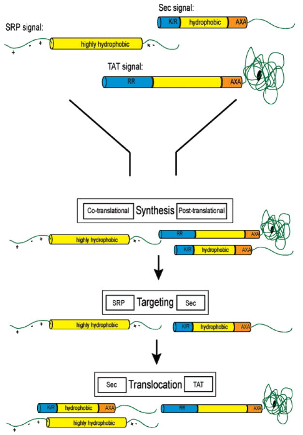
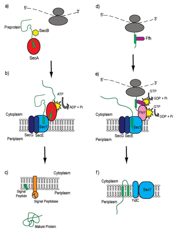
Understanding the transport of hydrophilic proteins across biological membranes continues to be an important undertaking. The general secretory (Sec) pathway in Escherichia coli transports the majority of E. coli proteins from their point of synthesis in the cytoplasm to their sites of final localization, associating sequentially with a number of protein components of the transport machinery. The targeting signals for these substrates must be discriminated from those of proteins transported via other pathways. While targeting signals for each route have common overall characteristics, individual signal peptides vary greatly in their amino acid sequences. How do these diverse signals interact specifically with the proteins that comprise the appropriate transport machinery and, at the same time, avoid targeting to an alternate route? The recent publication of the crystal structures of components of the Sec transport machinery now allows a more thorough consideration of the interactions of signal sequences with these components.


Figures

Figure 1
Figure 1
Classes of bacterial signal peptides. Sec signals are characterized by an amino-terminal positively charged domain of 5–6 residues, followed by a central hydrophobic core of 10–12 residues and a polar region of 6 residues containing the signal peptidase cleavage site. TAT signals contain slightly longer amino-terminal (containing the signature RR) and hydrophobic domains of 10–20 residues each, followed by a similar carboxyl-terminal cleavage region. SRP substrates are typically inner membrane proteins in which a long hydrophobic transmembrane domain acts as a signal anchor and is surrounded by polar regions that ultimately reside in the cytoplasm and periplasm. These signals are discriminated and funneled to the appropriate pathway based on these characteristics at various stages of the preproteins’ export. For example, the long hydrophobic domain of an SRP substrate is detected during synthesis; SRP associates cotranslationally with the nascent chain emerging from the ribosome and directs it to the SecYEG pore. Sec signals are detected posttranslationally and targeted by SecA to the SecYEG translocon. TAT signals are translocated posttranslationally via the TAT translocation channel. Blue, charged amino terminal region; yellow, hydrophobic region; orange, polar cleavage region.
Figure 2
Figure 2
Schematic representation of signal peptide interactions with components of the Sec machinery during transport. Signal peptides of secretory proteins are bound by SecA (a) in the cytoplasm, delivered to, bound by, and translocated via SecY of the pore (b), and ultimately bind to signal peptidase for cleavage of the signal from the mature protein (c). Inner membrane protein signals are bound by SRP as they emerge from the ribosome (d) and targeted to the membrane. The signal anchor interacts with SecY (e) and translocation occurs via the membrane-embedded SecYEG channel followed by membrane integration via YidC (f).
Figure 3
Figure 3
Models based on the crystal structures of Sec transport components with the predicted location of bound signal peptide (pink cylinder) illustrated: (A) Ffh from Thermus aquaticus (PDB ID 2FFH). Conserved residues that line the hydrophobic groove of the M-domain and are implicated in signal peptide binding are shown in blue. This region was entirely disordered in the E. coli structure (113) suggesting flexibility inherent in binding a variety of sequences. (B) SecA from Bacillus subtilis (PDB ID 1M6N). The residues proposed to bind signal peptide are shown in red (PPXD; 68), green (“stem” of PBD; 70), and blue (SPBG; 71). The signal peptide is shown as predicted in ref ; the SPBG overlaps with the PPXD and is adjacent to the PBD “stem”. (C) SecYEβ from Methanococcus jannaschii (PDB ID 1RHZ). Cross-linking data suggest that helices two and seven (shown in blue) interact with the signal peptide (93, 94). (D) Catalytic domain of E. coli signal peptidase (PDB ID 1KN9). Molecular modeling suggested hydrogen bonding of the signal peptide to β-sheets (shown in purple) that line the shallow substrate binding pocket (99).

References

    1. Engelman DM, Steitz TA. The spontaneous insertion of proteins into and across membranes: the helical hairpin hypothesis. Cell. 1981;23:411–422. - PubMed
    1. Luirink J, von Heijne G, Houben E, de Gier JW. Biogenesis of inner membrane proteins in Escherichia coli. Annu Rev Microbiol. 2005;59:329–355. - PubMed
    1. Chen M, Samuelson JC, Jiang F, Muller M, Kuhn A, Dalbey RE. Direct interaction of YidC with the Sec-independent Pf3 coat protein during its membrane protein insertion. J Biol Chem. 2002;277:7670–7675. - PubMed
    1. Sargent F, Berks BC, Palmer T. Pathfinders and trailblazers: a prokaryotic targeting system for transport of folded proteins. FEMS Microbiol Lett. 2006;254:198–207. - PubMed
    1. Blaudeck N, Kreutzenbeck P, Freudl R, Sprenger GA. Genetic analysis of pathway specificity during posttranslational protein translocation across the Escherichia coli plasma membrane. J Bacteriol. 2003;185:2811–2819. - PMC - PubMed

Publication types

MeSH terms