. 2007 Aug;5(8):e218.
doi: 10.1371/journal.pbio.0050218.

Tetrameric structure of centromeric nucleosomes in interphase Drosophila cells

Affiliations

Tetrameric structure of centromeric nucleosomes in interphase Drosophila cells

Yamini Dalal et al. PLoS Biol. 2007 Aug.

Abstract

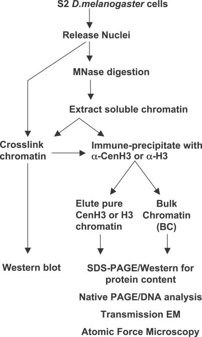
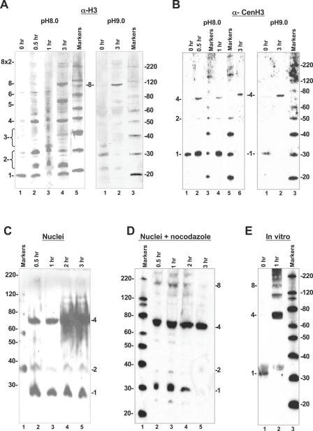
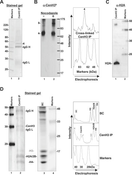
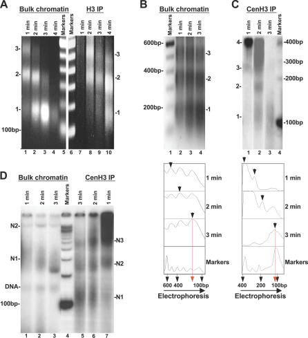
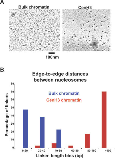
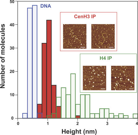
Centromeres, the specialized chromatin structures that are responsible for equal segregation of chromosomes at mitosis, are epigenetically maintained by a centromere-specific histone H3 variant (CenH3). However, the mechanistic basis for centromere maintenance is unknown. We investigated biochemical properties of CenH3 nucleosomes from Drosophila melanogaster cells. Cross-linking of CenH3 nucleosomes identifies heterotypic tetramers containing one copy of CenH3, H2A, H2B, and H4 each. Interphase CenH3 particles display a stable association of approximately 120 DNA base pairs. Purified centromeric nucleosomal arrays have typical "beads-on-a-string" appearance by electron microscopy but appear to resist condensation under physiological conditions. Atomic force microscopy reveals that native CenH3-containing nucleosomes are only half as high as canonical octameric nucleosomes are, confirming that the tetrameric structure detected by cross-linking comprises the entire interphase nucleosome particle. This demonstration of stable half-nucleosomes in vivo provides a possible basis for the instability of centromeric nucleosomes that are deposited in euchromatic regions, which might help maintain centromere identity.


Conflict of interest statement

Competing interests. The authors have declared that no competing interests exist.

Figures

Figure 1
Figure 1. Scheme to Analyze CenH3 Chromatin
D. melanogaster S2 or Kc cells were lysed gently with NP-40 to release nuclei. Nuclei were digested over a time course with MNase to release soluble chromatin extracted in 0.35 M salt buffer. Chromatin extracts and intact nuclei were used for protein cross-linking with 1 mg/ml DMS over a time course of 0–3 h. Western blot analysis of H3- and CenH3- nucleosomal complexes was performed on complexes resolved on gradient gels. Soluble chromatin extracts were used for immune-purification of native and in situ cross-linked CenH3-nucleosomes. Immunoprecipitations were subsequently analyzed by 18% SDS-PAGE, native PAGE, MNase digestion, and electron and atomic force microscopy.
Figure 2
Figure 2. Intranucleosomal Contacts Are Limited in CenH3 Nucleosomes
Western blots of D. melanogaster S2 chromatin extracts (A and B), S2 nuclei (C and D), and in vitro reconstituted CenH3 particles (E). Cross-linking was performed with 1 mg/ml DMS at pH 8.0 (left panels in A and B and panels C–E) or pH 9.0 (right panels in A and B) for the indicated times. Samples were resolved on 4%–12% NuPAGE or Tris-Glycine gradient gels. (A) Anti-H3 antibody detects enrichment of the limit octameric species over the cross-linking range in bulk chromatin extracts at pH 8.0 (left panel), and demonstrates enrichment of the octameric species at 110 kDa at the most cross-linked time point in lane 2 at pH 9.0 (right panel). Species annotated 1–8 refer to cross-linked monomer, dimer, and so on up to octamer. (B) Anti-CenH3 antibody probing duplicated samples (as in A) detects only two cross-linked products, deduced to be dimers and heterotypic tetramers (left panel, pH 8.0). Cross-linking to 3 h in pH 9.0 buffer demonstrates a conversion of most of the CenH3 to the species migrating at about 68 kDa. (C) Anti-CenH3 antibody detects robust amounts of a tetrameric species at approximately 68 kDa in cross-linked nuclei (lanes 2–5) obtained from asynchronous cells in culture. (D) Anti-CenH3 antibody detects predominantly tetrameric species at 68 kDa, as well as minor amounts of higher species (lanes 2–5) in cross-linked nuclei obtained from cells arrested at mitosis with nocodazole. (E) Anti-CenH3 antibody detects a cross-linking ladder of in vitro assembled salt-extracted native histones yielding robust amounts of species that are larger than tetramers (lane 2) in 1 h of cross-linking at pH 8.0. A similar range of products was obtained for in vitro assembled particles using recombinant CenH3 (Figure S1B).
Figure 3
Figure 3. CenH3 IP Particles Are Primarily Stable Heterotypic Tetramers at Interphase Containing H2A, H2B, and H4 in Approximately Equimolar Amounts
(A) A laser scan of SYPRO-Ruby stained 18% SDS-PAGE gel of cross-linked CenH3 IP particles shows a major species at 68 kDa. Similar formaldehyde cross-linked samples were analyzed by mass spectrometric analysis, and found to contain H2A, H2B and H4 (Table S1). IgG H and IgG L are heavy and light chain of the primary antibody used in the immunoprecipitation. Densitometric analysis to the right depicts an independent immunoprecipitation with similar result. Protein standard is Magic Marker, 20–220 kDa (Invitrogen, USA). (B) Western blot of cross-linked CenH3 IP particles probed with biotinylated anti-CenH3 antibody (α-CenH3b) detects heterotypic species 4 as the predominant species (lane 1). Nocodazole treatment of cells yields heterotypic species 4 as the dominant species, with the appearance of an octamer-size species (lane 2). Protein standard is pre-stained broad range marker 6.5–175 kDa (Invitrogen). (C) Detection of H2A in CenH3 IP particles. Formaldehyde cross-linked CenH3 IP particles (as in [B]) were boiled to reverse cross-links, and samples were electrophoresed on an 18% SDS-PAGE gel. The blot was probed with biotinylated anti-H2A antibody (Upstate catalog number 07–146). Protein standard is Magic Marker 20–220 kDa (Invitrogen). (D) CenH3 IP nucleosomes contain H2A, H2B, CenH3, and H4. A laser scan of a SYPRO-Ruby–stained 20% SDS-PAGE gel containing denatured CenH3 IP nucleosomes reveals approximately equimolar amounts of CenH3, H2A, H2B, and H4, and depletion of H3 (lane 2, and densitometric analysis to the far right), whereas the lane containing bulk chromatin (BC, lane 3, and densitometric analysis to the far right), resolved on the same gel, has normal core histone stoichiometry. Two protein standards were present on the gel: Magic Marker 20–220 kDa (Invitrogen) and pre-stained broad range Marker 6.5–175 kDa (Invitrogen).
Figure 4
Figure 4. Less DNA Is Protected from Nuclease Digestion of CenH3- Compared with H3-Nucleosomes
(A) Ethidium bromide stained 1.5% agarose gel showing DNA obtained from progressively MNase-digested bulk input chromatin (lanes 1–4) and control H3 IP chromatin (lanes 7–10). Both show core particle protection at 150 bp. Marker is 100-bp DNA ladder (Invitrogen). (B) Ethidium bromide stained gel showing purified DNA obtained from dilute MNase digestion of bulk chromatin input has a normal progression yielding a canonical MNase “ladder” and core protection of 150 bp. Marker is 100-bp DNA ladder (Invitrogen). The densitometric scan is presented below. (C) Autoradiogram showing purified DNA obtained from CenH3 immunoprecipitation of MNase-digested chromatin (lanes 1–3). DNA was radioactively end-labeled and resolved on 8% PAGE. The nucleosomal ladder appears relatively smeary and multiples of a unit repeat are not evident. Marker is 100-bp DNA ladder (Invitrogen). Densitometric analysis below demonstrates reduction in CenH3 core protection to approximately 100–120 bp rather than 150 bp seen for bulk chromatin (B). Lane 2 required a lower exposure than the rest of the gel. Marker is 100-bp DNA ladder. (D) Native PAGE of CenH3 IP nucleosomes obtained from successive time points of MNase-digested input (from [B], lanes 1–3) displays faster migration kinetics for CenH3 mononucleosomal species compared to BC species. Nucleosomes are radioactively end labeled in all lanes. Half of the gel containing CenH3 IP (lanes 5–7) needed a 5× longer exposure time than BC (lanes 1–3). The sharp band (DNA) is most likely free mononucleosomal DNA, and species N1, N2, and N3 refer to successive nucleosomal species. Marker is 100-bp DNA ladder.
Figure 5
Figure 5. CenH3 Arrays Display Native Chromatin “Beads on a String” Appearance with Unusual Condensation Behavior
(A) Nucleosomes of bulk chromatin are uniform-sized “beads” or particles with regular short linker DNA in low ionic strength buffer (left panel). Purified CenH3 IP nucleosomal arrays (>90%) display native chromatin's “beads on a string” appearance, but extended linker DNAs between CenH3 nucleosomes are suggestive of resistance to condensation under physiological salt conditions (right panel). The 100-nm bar was derived from magnification factors. (B) Edge-to-edge internucleosomal distances measured from two independent experiments using NIH Image J demonstrate that the more extended CenH3 linker DNAs are 2–3 times longer than bulk nucleosome linker lengths under comparable conditions.
Figure 6
Figure 6. CenH3 Nucleosomes Are Half the Height of Bulk Nucleosomes as Measured by AFM
Histogram depicting mean heights of control H4 IP nucleosomes (green bars) and CenH3 IP nucleosomes (red bars) computed from 100 counts of each. Mean H4 IP nucleosomal height is 2.05 ± 0.62 nm, in accordance with previously observed heights for bulk octameric nucleosomes. Mean CenH3 IP nucleosomal height is 1.03 ± 0.17 nm, or one-half octameric nucleosomal height, consistent with a tetrameric structure. Insets depict representative CenH3 IP molecules (inset, red box) and H4 IP molecules (inset, green box) used in this analysis. DNA heights (blue bars) were computed as an internal control for CenH3 IP nucleosomes. Scale bar represents 100 nm.

References

    1. Amor DJ, Kalitsis P, Sumer H, Choo KH. Building the centromere: From foundation proteins to 3D organization. Trends Cell Biol. 2004;14:359–368. - PubMed
    1. Malik HS, Henikoff S. Phylogenomics of the nucleosome. Nat Struct Biol. 2003;10:882–891. - PubMed
    1. Tomonaga T, Matsushita K, Yamaguchi S, Oohashi T, Shimada H, et al. Overexpression and mistargeting of centromere protein-A in human primary colorectal cancer. Cancer Res. 2003;63:3511–3516. - PubMed
    1. Ahmad K, Henikoff S. Histone H3 variants specify modes of chromatin assembly. Proc Natl Acad Sci U S A. 2002;99(Suppl 4):16477–16484. - PMC - PubMed
    1. Heun P, Erhardt S, Blower MD, Weiss S, Skora AD, et al. Mislocalization of the Drosophila centromere-specific histone CID promotes formation of functional ectopic kinetochores. Dev Cell. 2006;10:303–315. - PMC - PubMed

Publication types

MeSH terms