. 2007 Aug 2:7:129.
doi: 10.1186/1471-2148-7-129.

Molecular evolution of the MAGUK family in metazoan genomes

Affiliations

Molecular evolution of the MAGUK family in metazoan genomes

Aartjan J W te Velthuis et al. BMC Evol Biol. .

Abstract

Background: Development, differentiation and physiology of metazoans all depend on cell to cell communication and subsequent intracellular signal transduction. Often, these processes are orchestrated via sites of specialized cell-cell contact and involve receptors, adhesion molecules and scaffolding proteins. Several of these scaffolding proteins important for synaptic and cellular junctions belong to the large family of membrane-associated guanylate kinases (MAGUK). In order to elucidate the origin and the evolutionary history of the MAGUKs we investigated full-length cDNA, EST and genomic sequences of species in major phyla.

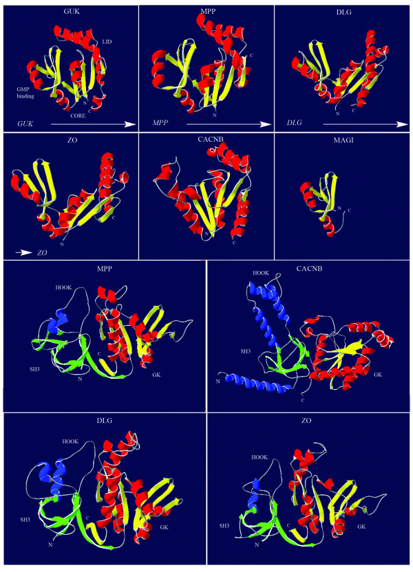
Results: Our results indicate that at least four of the seven MAGUK subfamilies were present in early metazoan lineages, such as Porifera. We employed domain sequence and structure based methods to infer a model for the evolutionary history of the MAGUKs. Notably, the phylogenetic trees for the guanylate kinase (GK)-, the PDZ- and the SH3-domains all suggested a matching evolutionary model which was further supported by molecular modeling of the 3D structures of different GK domains. We found no MAGUK in plants, fungi or other unicellular organisms, which suggests that the MAGUK core structure originated early in metazoan history.

Conclusion: In summary, we have characterized here the molecular and structural evolution of the large MAGUK family. Using the MAGUKs as an example, our results show that it is possible to derive a highly supported evolutionary model for important multidomain families by analyzing encoded protein domains. It further suggests that larger superfamilies encoded in the different genomes can be analyzed in a similar manner.


Figures

Figure 1
Figure 1
Domain architectures of the MAGUK subfamilies and their distribution over the eukaryotic phyla. (A) Eight subfamilies have been identified as members of the MAGUK family in our study. Depicted here in a general schematic representation are the domains present in these members. All members contain a central core comprised of a GK domain and several N and/or C-terminally positioned PDZ domains. All members except the MAGI proteins contain a SH3 domain that is nested between a PDZ and the GK domain. The CASK and MPP proteins contain N-terminal L27 domains, while CARMA genes encode CARD domains at this position. Names used are allowing a more systematic, but do not reflect in all cases the commonly used names e.g. DLG4 is better known as PSD95. A list of synonyms is given in table 1. The CACNB subfamily, commonly not regarded as a canonical MAGUK subfamily, only contains the SH3 and GK domains. Phylogenetic analysis presented here however shows that it is a related subfamily. (B) The MAGUK subfamily distribution over the eukaryotic phyla shows no homologs were found for Choanozoans/Protozoans, Plantae and Fungi, here represented by Monosiga ovata, Arabidopsis thaliana and Saccharomyces cerevisae, respectively. Tetraodon nigroviridis has duplicated DLG4, ZO1 and ZO2 encoding genes, whereas Gallus gallus lacked the gene for DLG4. The arrowheads on the bottom indicate two possible gene duplication events.
Figure 2
Figure 2
Guanylate kinase dendrogram and summarized phylogenetic analyses. (A) The dendrogram is based on all protein families currently known to contain GK sequences (note: this is not a phylogeny). These include the three families of the voltage-gated calcium channel beta subunit (CAB), the homologs of Guanylate kinase (GUK) and the MAGUK family. The core structure of the GUK is GK only, while the CAB and MAGUK families have a SH3-GK or PDZ-SH3-GK architecture respectively. The MAGUK and CAB families were only found in metazoan species, while sequences of GUK family members appeared dispersed over all eukaryotic lineages. In the GUK clade the metazoans are indicated with the letter M, the Fungi with F, Bacteria with B, Viruses with V and the Plants with P. Species that were included in this dendrogram as well as sequences used are listed in Additional file 2. (B-C) Summarized phylogenetic trees based on Bayesian consensus trees (see Additional file 3, 4, 5) for the GK, SH3 and PDZ domains, respectively. Numbers indicate % Bayesian posterior probability and % bootstrap Maximum Likelihood.
Figure 3
Figure 3
Molecular modeling of guanylate kinase domains based on the human sequences. Indicated in the left, uppermost panel are the three dynamic domains of the GMP guanylate Kinase: the GMP binding sub-domain, the CORE sub-domain and the LID sub-domain. The other panels show modeled structures of the MAGUK subfamilies, superimposed by Swiss Model. Arrows indicate the likely course of the structural evolution. The relationships established through our phylogenetic analysis are reflected here in the comparison of structures for the human representatives. The MPP structure still has a resemblance close to the human GMP guanylate Kinase, having a similar orientation of the three sub-domains of the GMP GK. The DLG and ZO subfamilies are also very closely related, when the presented 3D structures are compared. The MAGI GK could only be partially superimposed on known structures, sharing at least the GK GMP binding domain with the other MAGUK subfamilies. The used PDB files are listed in Additional file 2. The lowest four panels show representative models of the GK-HOOK-SH3 super domains of the MAGUK subfamilies MPP, DLG, ZO and CACNB. As in the upper panels the GK domain is colored yellow and magenta (β-strands and helices respectively), while the SH3 and HOOK regions are colored green and blue.
Figure 4
Figure 4
Evolutionary model of the MAGUK subfamily-structure evolution. Evolving from a catalytically active GK encoding gene, the family arose by obtaining both a PDZ and SH3 domain. Then the MPP subfamily branched off, assimilating L27 domains and, in the case of CASK, also a kinase domain. The MPP subfamily member known as MPP1 evolved last and lost its L27 domains. The CACNB and MAGI subfamilies arose through domain loss of the PDZ and SH3 domains, respectively. The later one also obtained WW domains, most likely resulting in a C-terminal truncation of the GK domain. The MAGUK core structure, consisting of a PDZ, SH3 and GK domain evolved further and gave rise, after duplication of the most C-terminal PDZ domain, to the DLG, ZO and, lastly, the CARMA subfamily. This PDZ domain duplicated another time at the birth of the DLG5 structure. The arrowheads on the bottom indicate two different gene duplication events. Arrows on top of the protein structures show PDZ domain duplication events derived from our phylogeny.

References

    1. Funke L, Dakoji S, Bredt DS. Membrane-associated Guanylate Kinases Regulate Adhesion and Plasticity at Cell Junctions. Annual Review of Biochemistry. 2005;74:219–245. doi: 10.1146/annurev.biochem.74.082803.133339. - DOI - PubMed
    1. González-Mariscal L. Betanzos A. Ávila-Flores A. MAGUK proteins: structure and role in the tight junction. Seminars in Cell & Developmental Biology. 2002;11:315–324. doi: 10.1006/scdb.2000.0178. - DOI - PubMed
    1. Mburu P, Kikkawa Y, Townsend S, Romero R, Yonekawa H, Brown SDM. Whirlin complexes with p55 at the stereocilia tip during hair cell development. PNAS. 2006;103:10973–10978. doi: 10.1073/pnas.0600923103. - DOI - PMC - PubMed
    1. Elias EM, Funke L, Stein V, Grant SG, Bredt DS, Nicoll RA. Synapse-Specific and Developmentally Regulated Targeting of AMPA Receptors by a Family of MAGUK Scaffolding Proteins . Neuron. 2006;52:307–320. doi: 10.1016/j.neuron.2006.09.012. - DOI - PubMed
    1. Kornau HC, Schenker LT, Kennedy MB, Seeburg PH. Domain interaction between NMDA receptor subunits and the postsynaptic density protein PSD-95. Science. 1995;269:1737–1740. doi: 10.1126/science.7569905. - DOI - PubMed