. 2008 Jan 1;314(1):11-24.
doi: 10.1016/j.yexcr.2007.06.007. Epub 2007 Jun 27.

Tissue inhibitor of metalloproteinase-2 (TIMP-2) regulates myogenesis and beta1 integrin expression in vitro

Affiliations

Tissue inhibitor of metalloproteinase-2 (TIMP-2) regulates myogenesis and beta1 integrin expression in vitro

Gentian Lluri et al. Exp Cell Res. .

Abstract

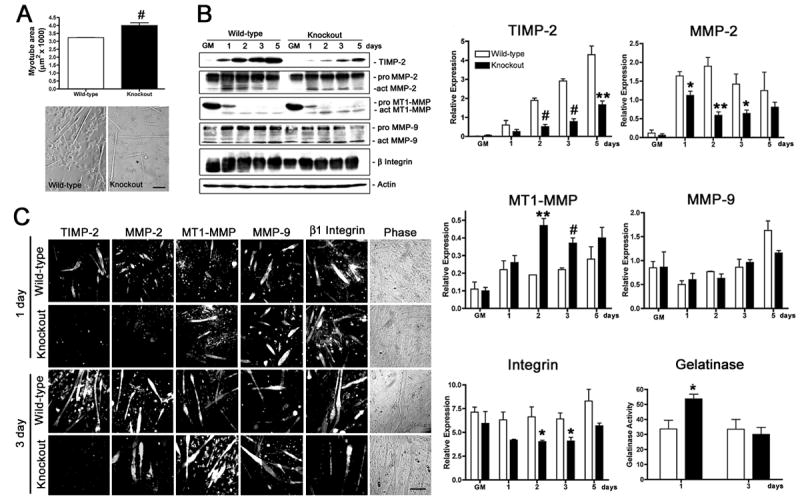
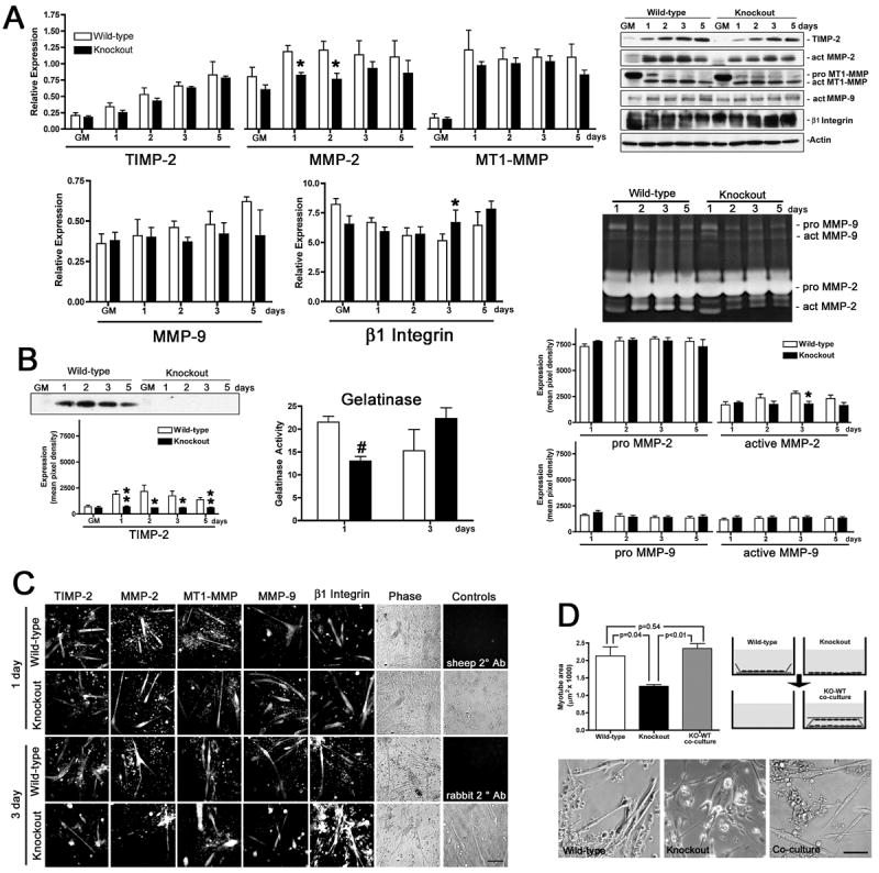
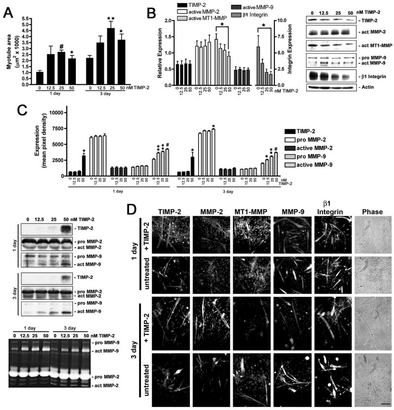
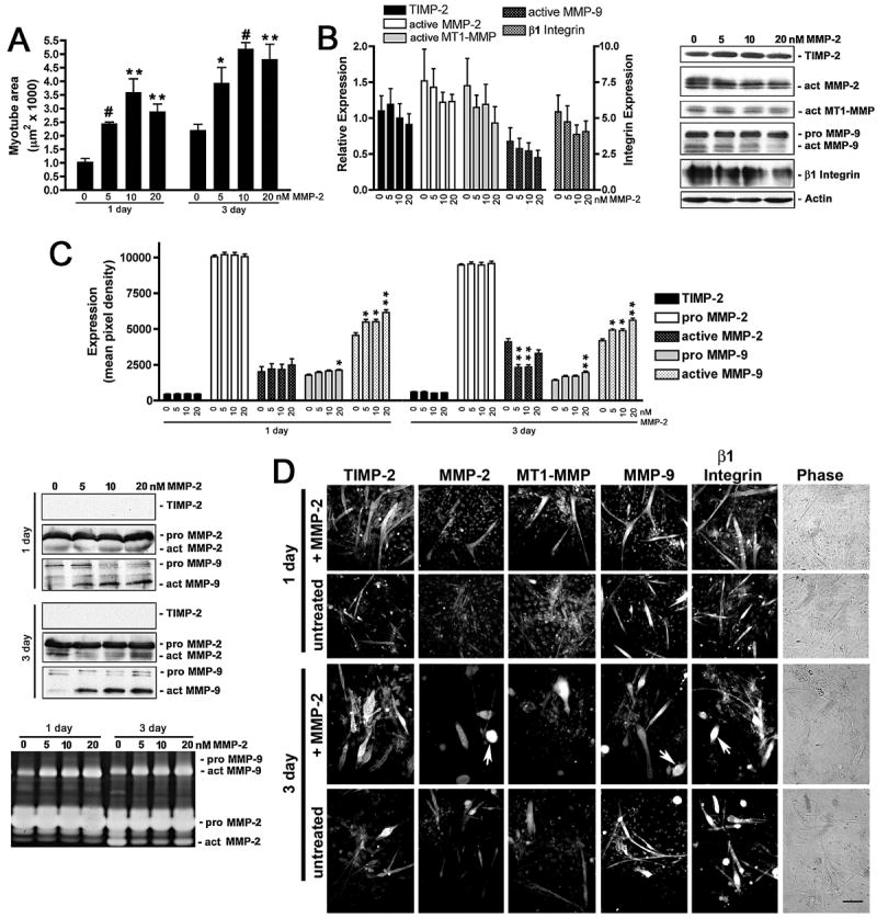
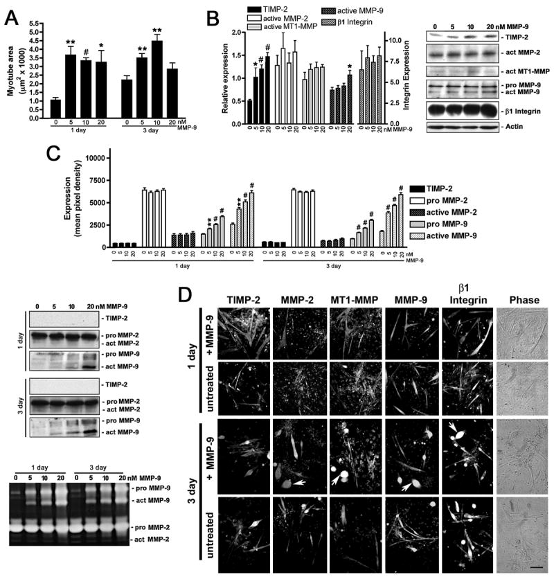
Myogenesis in vitro involves myoblast cell cycle arrest, migration, and fusion to form multinucleated myotubes. Extracellular matrix (ECM) integrity during these processes is maintained by the opposing actions of matrix metalloproteinase (MMP) proteases and their inhibitors, the tissue inhibitor of metalloproteinases (TIMPs). Here, we report that TIMP-2, MMP-2, and MT1-MMP are differentially expressed during mouse myoblast differentiation in vitro. A specific role for TIMP-2 in myogenesis is demonstrated by altered TIMP-2(-/-) myotube formation. When differentiated in horse serum-containing medium, TIMP-2(-/-) myotubes are larger than wild-type myotubes. In contrast, when serum-free medium is used, TIMP-2(-/-) myotubes are smaller than wild-type myotubes. Regardless of culture condition, myotube size is directly correlated with MMP activity and inversely correlated with beta1 integrin expression. Treatment with recombinant TIMP-2 rescues reduced TIMP-2(-/-) myotube size and induces increased MMP-9 activation and decreased beta1 integrin expression. Treatment with either MMP-2 or MMP-9 similarly rescues reduced myotube size, but has no effect on beta1 integrin expression. These data suggest a specific regulatory relationship between TIMP-2 and beta1 integrin during myogenesis. Elucidating the role of TIMP-2 in myogenesis in vitro may lead to new therapeutic options for the use of TIMP-2 in myopathies and muscular dystrophies in vivo.


Figures

Figure 1
Figure 1. TIMP-2−/− myotubes are larger than wild-type myotubes when cultured in the presence of horse serum
A) After 3 days in differentiation media containing 2% heat-inactivated horse serum, cells were fixed, and myotube area was measured (at least 20 randomly selected myotubes were measured from four wild-type and three TIMP-2−/− cultures). Data are presented as mean ± s.e.m. TIMP-2−/− myotubes are larger than wild-type myotubes. Myonuclei number is also increased in TIMP-2−/− myotubes, suggesting increased fusion (not shown). B) Western blot analysis with whole cell lysates (25 μg protein) of rapidly proliferating cells maintained in growth media (GM) for 24 hours after plating and after 1, 2, 3, and 5 days in differentiation media containing 2% heat-inactivated horse serum. Densitometric analysis (normalized to actin at each time point) reveals a dramatic reduction in TIMP-2 and active MMP-2 expression, increased active MT1-MMP expression, and no change in active MMP-9 expression in TIMP-2−/− myotubes. Similar to TIMP-2−/− muscle in vivo, total cellular β1 integrin expression, as well as cell surface expression (not shown), is decreased in TIMP-2−/− myotubes at 3 days, when myotube size is increased. Using a fluorescently caged gelatin substrate (i.e., DQ gelatin), protease activity was measured in 100 μl conditioned media (normalized to unconditioned medium containing serum). No difference in net gelatinolytic activity is detected at 3 days; in contrast, gelatinolytic activity is significantly increased at 1 day, the period of myoblast migration and fusion. Data are presented as mean ± s.e.m. C) Immunocytochemistry of Triton permeabilized cells confirms the western blot data of decreased TIMP-2 and MMP-2 expression and increased MT1-MMP expression in TIMP-2−/− myotubes. *p < 0.05, **p ≤ 0.01, #p ≤ 0.001. Scale bar = 100 μm.
Figure 2
Figure 2. TIMP-2−/− myotubes are smaller than wild-type myotubes when cultured in serum-free media
A) Western blot analysis (25 μg whole cell lysates) of myotubes differentiated in serum-free medium reveals that the protein expression profile and developmental regulation differs little between wild-type and TIMP-2−/− myotubes. Notable exceptions include decreased active MMP-2 expression at 1 and 2 days and increased β1 integrin expression at 3 days. B) Analysis of conditioned media. Western blot analysis (50 μl conditioned medium) shows that although TIMP-2−/− myotubes possess TIMP-2 protein (Fig. 1B) it is not secreted, indicating a biological knockout for extracellular TIMP-2 activity (i.e., MMP inhibition and proMMP-2 activation). Gelatinase activity (in 100 μl conditioned differentiation medium normalized to unconditioned media containing N2 supplement) is decreased at 1 day, but unchanged at 3days. By zymography (50 μl conditioned medium), decreased active MMP-2 expression is detected at 3 days, but not at 1 day, indicating that MMPs other than MMP-2 and MMP-9 contribute to the decreased net gelatinolytic activity. C) Immunocytochemistry of Triton permeabilized cells confirms the western blot data and reveals that both wild-type and TIMP-2−/− cultures contain a greater number of undifferentiated cells relative to cultures differentiated with horse serum. Secondary antibody alone controls (sheep and rabbit 2° Ab) demonstrate the specificity of immunolabeling. D) After 3 days in serum-free differentiation medium, cells were fixed and myotube area was measured. In contrast to myotubes differentiated with 2% horse serum (Fig. 1), TIMP-2−/− myotubes in serum-free media are smaller than wild-type myotubes. Co-culture of TIMP-2−/− (KO) with wild-type (WT) myotubes in close, but not direct, contact increases myotube size comparable to wild-type levels, suggesting soluble factor(s) are responsible for the rescue. Photomicrographs representative of the three culture conditions are shown. *p < 0.05, **p ≤ 0.01, #p ≤ 0.001. Scale bars = 100 μm.
Figure 3
Figure 3. TIMP-2 rescues reduced TIMP-2−/− myotube size, increases active MMP-9, and decreases β1 integrin expression
A) TIMP-2−/− myotube area was measured on live cells after 1 and 3 days in differentiation media in the absence or presence of recombinant active mouse TIMP-2 (12.5, 25, or 50 nM). Both at 1 and 3 days, 25 nM TIMP-2 is most effective at rescuing the reduced TIMP-2−/− myotube size. B) Western blot analysis (25 μg whole cell lysates) shows decreased active MT1-MMP and β1 integrin expression in TIMP-2 treated cultures at 3 days. C) Western blot and zymographic analysis (25 μl conditioned medium) reveals a dose-dependent increase in active MMP-9 secretion in response to TIMP-2 treatment both at 1 and 3 days. D) Immunocytochemistry of Triton permeabilized untreated and TIMP-2 treated (25 nM) cells. *p < 0.05, **p ≤ 0.01, #p ≤ 0.001. Scale bar = 100 μm.
Figure 4
Figure 4. MMP-2 rescues reduced TIMP-2−/− myotube size and increases active MMP-9 without altering β1 integrin expression
A) TIMP-2−/− myotube area was measured on live cells after 1 and 3 days in differentiation media in the absence or presence of recombinant active mouse MMP-2 (5, 10, 20 nM). Both at 1 and 3 days, 10 nM MMP-2 is most effective at rescuing the reduced TIMP-2−/− myotube size. B) Western blot analysis (25 μg whole cell lysates) shows that MMP-2 treatment has no effect on the proteins analyzed. C) Western blot and zymographic analysis (25 μl conditioned medium) reveals a dose-dependent increase in active MMP-9 secretion in response to MMP-2 treatment both at 1 and 3 days. In addition, MMP-2 treatment (5 and 10 nM) decreases active MMP-2 secretion at 3 days. D) Immunocytochemistry of Triton permeabilized untreated and MMP-2 treated (10 nM) cells. Note that the number of myoballs (arrows) is increased in MMP-2 treated cultures at 3 days. *p < 0.05, **p ≤ 0.01, #p ≤ 0.001. Scale bar = 100 μm.
Figure 5
Figure 5. MMP-9 rescues reduced TIMP-2−/− myotube size without altering β1 integrin expression
A) TIMP-2−/− myotube area was measured on live cells after 1 and 3 days in differentiation media in the absence or presence of recombinant active mouse MMP-9 (5, 10, 20 nM). MMP-9 rescues TIMP-2−/− myotube size at a lower concentration (5 nM) than MMP-2 (10 nM) at both 1 and 3 days. B) Western blot analysis (25 μg whole cell lysates) shows that MMP-9 treatment increases cellular TIMP-2 expression, but has no effect on β1 integrin expression. C) Western blot and zymographic analysis (25 μl conditioned medium) detects increased active MMP-9 in the medium of MMP-9 treated cells. However, no change in TIMP-2 secretion is detected. D) Immunocytochemistry of Triton permeabilized untreated and MMP-9 treated (10 nM) cells. Note that, like MMP-2 treatment, the number of myoballs (arrows) is increased in MMP-9 treated cultures at 3 days. *p < 0.05, **p ≤ 0.01, #p ≤ 0.001. Scale bar = 100 μm.

Similar articles

Cited by

References

    1. Parker MH, Seale P, Rudnicki MA. Looking back to the embryo: defining transcriptional networks in adult myogenesis. Nat Rev Genet. 2003;4:497–507. - PubMed
    1. Brand-Saberi B. Genetic and epigenetic control of skeletal muscle development. Ann Anat. 2005;187:199–207. - PubMed
    1. Krauss RS, Cole F, Gaio U, Takaesu G, Zhang W, Kang JS. Close encounters: regulation of vertebrate skeletal myogenesis by cell-cell contact. J Cell Sci. 2005;118:2355–2362. - PubMed
    1. Nagase H, Woessner JF. Matrix metalloproteinases. J Biol Chem. 1999;274:21491–21494. - PubMed
    1. McCawley LJ, Matrisian LM. Matrix metalloproteinases: they’re not just for matrix anymore! Curr Opin Cell Biol. 2001;13:534–540. - PubMed

Publication types

MeSH terms

Substances