. 2007 Aug 3;27(3):462-73.
doi: 10.1016/j.molcel.2007.06.013.

APC/C(Cdc20) controls the ubiquitin-mediated degradation of p21 in prometaphase

Affiliations

APC/C(Cdc20) controls the ubiquitin-mediated degradation of p21 in prometaphase

Virginia Amador et al. Mol Cell. .

Abstract

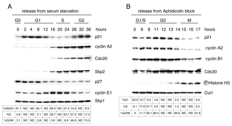
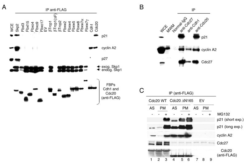
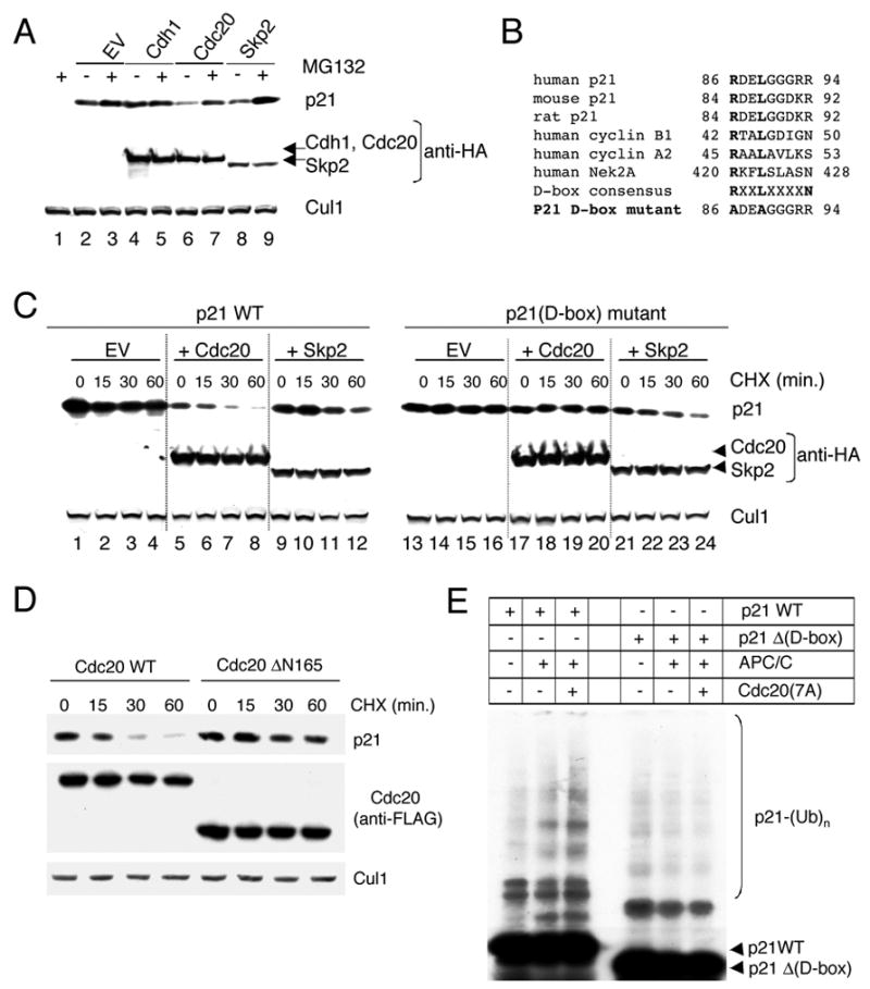
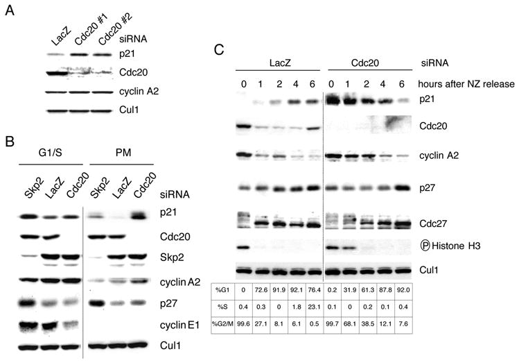
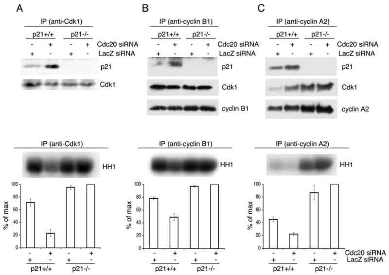
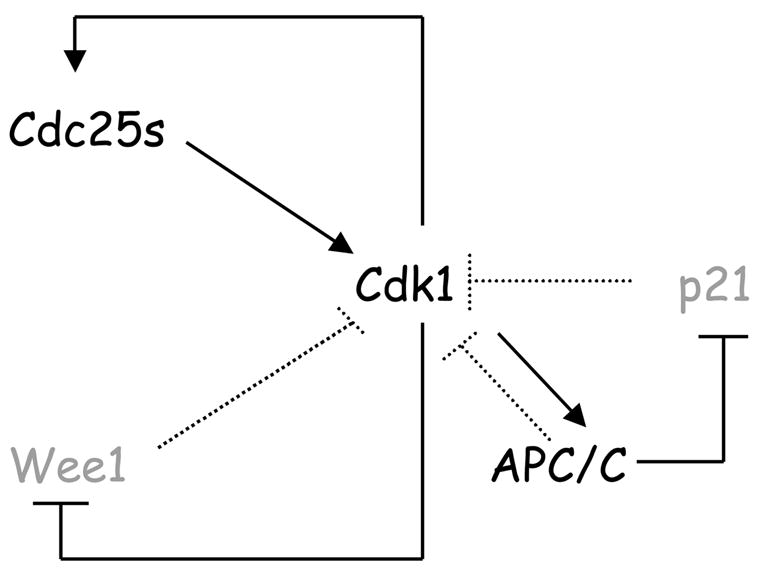
During the G1/S transition, p21 proteolysis is mediated by Skp2; however, p21 reaccumulates in G2 and is degraded again in prometaphase. How p21 degradation is controlled in mitosis remains unexplored. We found that Cdc20 (an activator of the ubiquitin ligase APC/C) binds p21 in cultured cells and identified a D box motif in p21 necessary for APC/C(Cdc20)-mediated ubiquitylation of p21. Overexpression of Cdc20 or Skp2 destabilized wild-type p21; however, only Skp2, but not Cdc20, was able to destabilize a p21(D box) mutant. Silencing of Cdc20 induced an accumulation of p21, increased the fraction of p21 bound to Cdk1, and inhibited Cdk1 activity in p21(+/+) prometaphase cells, but not in p21(-/-) cells. Thus, in prometaphase Cdc20 positively regulates Cdk1 by mediating the degradation of p21. We propose that the APC/C(Cdc20)-mediated degradation of p21 contributes to the full activation of Cdk1 necessary for mitotic events and prevents mitotic slippage during spindle checkpoint activation.


Figures

Figure 1
Figure 1. p21 is degraded in early mitosis
(A) T98G cells were synchronized in G0 by 72 hours of serum starvation (indicated as time 0). Cells were then re-stimulated with serum and collected at the indicated time points. Protein extracts were analyzed by immunobloting with antibodies against the indicated proteins. Cell cycle phases were monitored by flow cytometry. (B) IMR-90 fibroblasts were first synchronized in G0 by 72 hours of serum starvation and then at the G1/S boundary (indicated as time 0) by re-stimulating them with serum for 28 hours, adding aphidicolin for the last 18 hours. Cells were subsequently washed and allowed to progress thorough the cell cycle for the indicated times. Protein extracts were analyzed by immunoblotting with antibodies against the indicated proteins. Cell cycle phases were monitored by flow cytometry.
Figure 2
Figure 2. Mitotic degradation of p21 is not dependent on Skp2
Mouse embryonic fibroblasts from Skp2−/ −;p27+/+, Skp2−/ −;p27−/ −, and Skp2+/+;p27+/+ mice were synchronized at G1/S by a double thymidine block (S-phase). Cells were subsequently washed and allowed to progress through the cell cycle for 12 hours in the presence of nocodazole. The prometaphase, round cells were collected by gentle shake-off, and finally, the remaining attached cells (G2-phase) were also harvested. Protein extracts were analyzed by immunobloting with antibodies to the indicated proteins.
Figure 3
Figure 3. p21 interacts with Cdc20
(A) HEK293T cells were transfected with an empty vector (EV) or constructs encoding the indicated FLAG–tagged F-box proteins (FBPs), FLAG-Cdh1, or FLAG-Cdc20, all in the presence of a plasmid encoding His-tagged Skp1. During the last six hours before harvesting, cells were treated with the proteasome inhibitor MG132. Exogenous proteins were immunoprecipitated (IP) from whole cell extracts (WCE) with an anti-FLAG resin, and immunocomplexes were probed with antibodies to the indicated proteins. (B) Endogenous Cdc20 and Cdc27 co-immunoprecipitates endogenous p21. Whole cell extracts (WCE) from HeLa cells were immunoprecipitated (IP) with normal IgG, anti-Cdc20, anti-Cdc27, or anti-Cdh1 antibodies. Immunocomplexes were probed with antibodies to the indicated proteins. The second lane contained molecular weight markers (MWM) that appear to cross-react with the anti-cyclin A2 antibody. (C) HeLa cells were infected with an empty vector (EV) or constructs encoding FLAG–tagged Cdc20 or FLAG–tagged Cdc20 Δ165 mutant. Where indicated, the proteasome inhibitor MG132 was added during the last six hours in culture. Exogenous proteins were immunoprecipitated (IP) from extracts obtained from asynchronous (AS) or prometaphase (PM) cells using an anti-FLAG resin. Immunocomplexes were then probed with antibodies to the indicated proteins.
Figure 4
Figure 4. Cdc20-dependent ubiquitylation and degradation of p21 depends on the D-box of p21
(A) Whole cell extracts were prepared from HEK293T cells transfected with p21 and either HA-tagged Cdc20, HA-tagged Cdh1, or HA-tagged Skp2. Levels of p21 were detected by immunoblotting with an anti-p21 antibody, whereas Cdc20, Cdh1, and Skp2 were analyzed using an anti-HA antibody. Cul1 was used as a loading control. Where indicated, the proteasome inhibitor MG132 was added during the last six hours in culture. No significant changes in the cell cycle phases (as monitored by flow cytometry) of transfected HEK293T cells were observed compared to controls. (B) Alignment of the amino acid regions corresponding to the putative Destruction-box motif (D-box) in human, mouse, and rat p21 with the D-box motifs of human cyclin B1, cyclin A2 and Nek2A. A schematic representation of the Arg- and Leu-to-Ala substitutions in the putative D-box motif of human p21 is depicted at the bottom. (C) HEK293T cells were transfected with wild type p21 or the p21(D-box) mutant in the presence of either an empty vector (EV), HA-tagged Cdc20, or HA-tagged Skp2. Twenty-four hours after transfection, cells were treated with cycloheximide (CHX) and the half-life of p21 analyzed by immunobloting. (D) HEK293T cells were transfected with wild type p21 in the presence of either FLAG–tagged Cdc20 or FLAG–tagged Cdc20 Δ165 mutant. Twenty-four hours after transfection, cells were treated with cycloheximide (CHX), and the half-life of p21 was analyzed by immunobloting. (E) In vitro ubiquitin ligation assay of 35S labeled in vitro transcribed/translated p21 or p21Δ (D-box) mutant was conducted in the absence or presence of immunopurified APC/C and constitutively active Cdc20(7A) mutant, as indicated. Samples were incubated at 30°C for 90 minutes. The bracket on the right side of the panels marks a ladder of bands corresponding to polyubiquitylated p21.
Figure 5
Figure 5. Cdc20 targets p21 for degradation in prometaphase
(A) HeLa cells were transfected twice with siRNA molecules to a non-relevant mRNA (Lac Z) or to Cdc20 mRNA (the latter using two different siRNA oligos: #1 and #2). Protein extracts were probed with antibodies to the indicated proteins. (B) U-2 OS cells were transfected twice with siRNA oligos to a non-relevant mRNA (Lac Z) or to Cdc20 mRNA. Cells were then treated with aphidicolin for 24 hours to arrest cells at G1/S (first 3 lanes). In a parallel experiment, aphidicolin was washed away to allow cells to progress through the cell cycle for an additional 18 hours in the presence of nocodazole (last 3 lanes). The prometaphase (PM), round cells were collected by gentle shake-off. Cell extracts were subjected to immunobloting with antibodies to the indicated proteins. (C) U-2 OS cells were transfected twice with siRNA oligos corresponding to a non-relevant mRNA (Lac Z) or to Cdc20 mRNA. Twelve hours after the last transfection, nocodazole (NZ) was added for an additional sixteen hours. Round, prometaphase cells were then collected by gentle shake-off and replated in fresh medium for the indicated times. Cells were harvested, and cell extracts were analyzed by immunoblotting with antibodies to the indicated proteins. Cell cycle phases were monitored by flow cytometry.
Figure 6
Figure 6. Cdc20-mediated degradation of p21 activates Cdk1 in early prometaphase
Hct116 p21 +/+ and Hct116 p21 –/−cells were transfected twice with siRNA oligos to a non-relevant mRNA (Lac Z) or to Cdc20 mRNA, as indicated. Cells were then synchronized at G1/S using a double thymidine block and then released to allow cells to progress through the cell cycle for 18 hours (adding nocodazole during the last 15 hours). Prometaphase cells were collected by gentle shake-off. Cell extracts were subjected to immunoprecipitation (IP) with antibodies against Cdk1 (A), cyclin B1 (B), or cyclin A2 (C). Immunocomplexes were probed with antibodies to the indicated proteins (upper panels) or used to perform kinase assays using Histone H1 (HH1) as a substrate (bottom panels). The error bars represent +/−SD from triplicate experiments quantified by measuring radioactivity in the HH1 band excised from the dried gel. A representative experiment showing an autoradiograph of 32P-labeled HH1 is presented on the top of the bottom panels.
Figure 7
Figure 7. Model of Cdk1 autoactivation via three feedback loops, including the activation of the APC/CCdc20-mediated degradation of p21 occurring in early mitosis
Black signifies activated forms of the respective proteins, gray indicates inactive forms or degraded proteins. Solid lines indicate activating (→) or inhibitory (--I) activities in prometaphase, dotted lines indicate activating or inhibitory activities in different phase of the cell cycle. See text for details.

Similar articles

Cited by

References

    1. Acquaviva C, Pines J. The anaphase-promoting complex/cyclosome: APC/C. J Cell Sci. 2006;119:2401–2404. - PubMed
    1. Bashir T, Dorrello NV, Amador V, Guardavaccaro D, Pagano M. Control of the SCF(Skp2-Cks1) ubiquitin ligase by the APC/C(Cdh1) ubiquitin ligase. Nature. 2004;428:190–193. - PubMed
    1. Bates S, Ryan KM, Phillips AC, Vousden KH. Cell cycle arrest and DNA endoreduplication following p21Waf1/Cip1 expression. Oncogene. 1998;17:1691–1703. - PubMed
    1. Bloom J, Amador V, Bartolini F, DeMartino G, Pagano M. Proteasome-Mediated Degradation of p21 via N-Terminal Ubiquitinylation. Cell. 2003;115:71–82. - PubMed
    1. Bloom J, Pagano M. To be or not to be ubiquitinated? Cell Cycle. 2004;3:138–140. - PubMed

Publication types

MeSH terms

LinkOut - more resources