. 2007 Aug 3;27(3):486-97.
doi: 10.1016/j.molcel.2007.06.015.

A phosphatase activity of Sts-1 contributes to the suppression of TCR signaling

Affiliations

A phosphatase activity of Sts-1 contributes to the suppression of TCR signaling

Anatoly Mikhailik et al. Mol Cell. .

Abstract

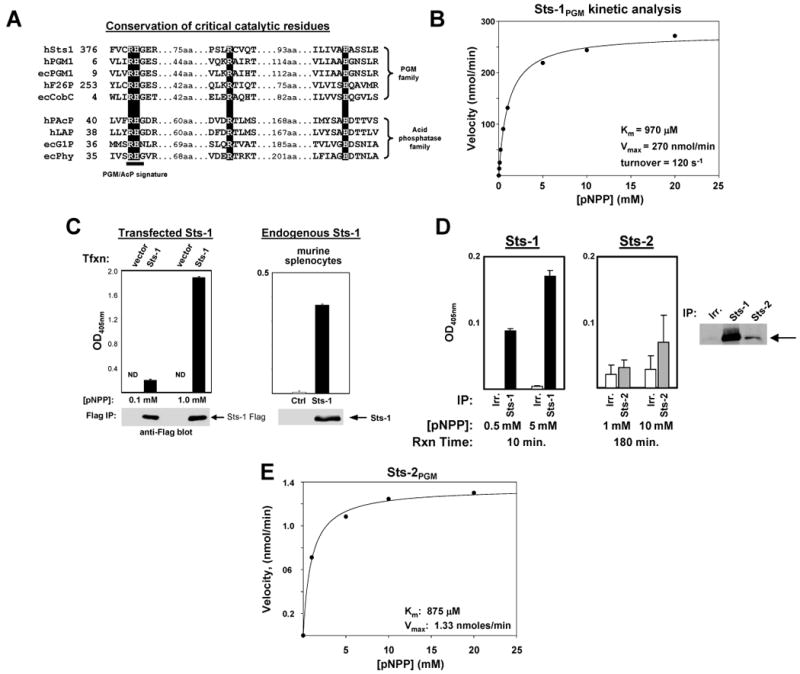
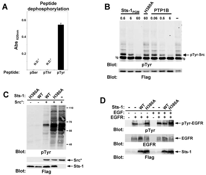
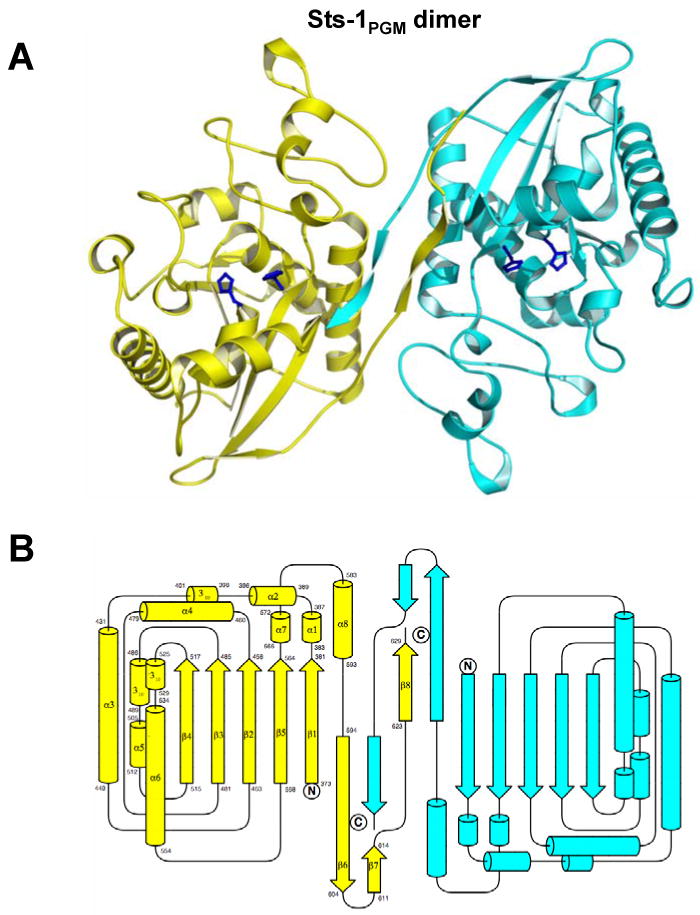
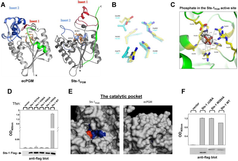
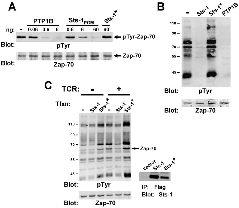
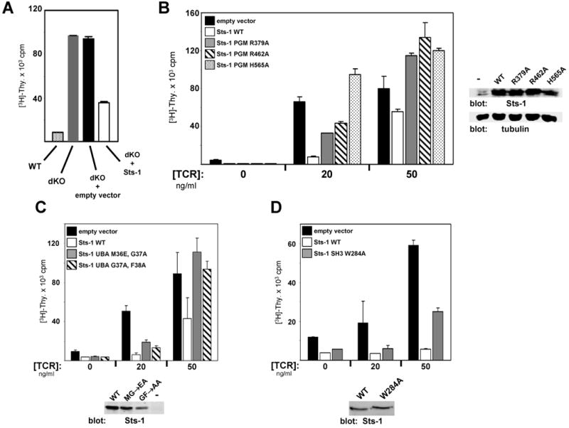
Precise signaling by the T cell receptor (TCR) is crucial for a proper immune response. To ensure that T cells respond appropriately to antigenic stimuli, TCR signaling pathways are subject to multiple levels of regulation. Sts-1 negatively regulates signaling pathways downstream of the TCR by an unknown mechanism(s). Here, we demonstrate that Sts-1 is a phosphatase that can target the tyrosine kinase Zap-70 among other proteins. The X-ray structure of the Sts-1 C terminus reveals that it has homology to members of the phosphoglycerate mutase/acid phosphatase (PGM/AcP) family of enzymes, with residues known to be important for PGM/AcP catalytic activity conserved in nature and position in Sts-1. Point mutations that impair Sts-1 phosphatase activity in vitro also impair the ability of Sts-1 to regulate TCR signaling in T cells. These observations reveal a PGM/AcP-like enzyme activity involved in the control of antigen receptor signaling.


Figures

Figure 1
Figure 1. Phosphatase activity of Sts-1
(A) Conservation of active site residues. The alignment program CLUSTALW (Chenna et. al., 2003) was used to align the Sts-1 C-terminus with PGM/AcP family members: hPGM1, human phosphoglycerate mutase isoform 1; hF26P, the phosphatase domain of human 6-phosphofructo-2-kinase/fructose-2,6-bisphosphatase; ecCobC, E. coli alpha ribazole phosphatase; hPAcP, human prostatic acid phosphatase; hLAP, human lysosomal acid phosphatase; ecG1P, E. coli glucose-1-phosphatase; ecPhy, E. coli phytase. A solid bar indicates the PGM/AcP signature motif, and the conserved active site arginines and histidines are shaded. (B) Kinetic analysis of Sts-1PGM catalytic activity. The velocity of pNPP hydrolysis by Sts-1PGM was at multiple substrate concentrations. Lineweaver-Burke analysis was used to calculated Km and Vmax values. (C) (left) Full-length Sts-1 has an associated phosphatase activity. Flag-tagged Sts-1 or control vectors were expressed in 293T cells, precipitated with anti-flag M2, and evaluated for pNPP phosphatase activity at the indicated substrate concentrations. The level of product was determined by measuring the OD405nm of the reaction supernatants. Each assay was conducted in triplicate (see error bars) and data representative of three separate experiments is displayed. Representative levels of Sts-1 in the reactions, as evaluated by SDS-PAGE, are illustrated. ND = not detectable. (right) Phosphatase activity of endogenous Sts-1. Activity assays were conducted on Sts-1 that was precipitated from murine splenocytes using Sts-1 specific antibodies. Analysis was conducted as described for transfected Sts-1. (D) Difference in pNPP hydrolysis activity between Sts-1 and Sts-2. Assays were conducted in triplicate (error bars) with two different concentrations of pNPP, using Sts proteins precipitated from Jurkat cells. Representative levels of Sts-1 or -2 in the reactions are illustrated. Note different reaction times. (E) Analysis of recombinant Sts-2PGM catalytic activity. The velocity of pNPP hydrolysis by Sts-2PGM (250 nM) was evaluated at multiple substrate concentrations. Lineweaver-Burke analysis was used to calculated Km and Vmax values.
Figure 2
Figure 2. Phosphotyrosine phosphatase activity of Sts-1
(A) Peptide dephosphorylation by Sts-1PGM. Sts-1PGM was tested for its ability to dephosphorylate peptides (500 μM) containing pSer and pThr (RRApSVA and KRpTIRR, UBI, Inc.), and pTyr (EIYAA-PFAKKGGGEpYENP). Levels of free phosphate were determined using malachite green. Error bars as in Figure 1. (B) Dephosphorylation of a tyrosine phosphorylated protein by Sts-1PGM. Dose dependent protein tyrosine phosphatase activity of Sts-1PGM, as compared to PTP1B, is illustrated. Constitutively active Flag-tagged Src was expressed in 293T cells, isolated by precipitation with anti-flag M2 antibodies, and utilized as a substrate. Reaction products were evaluated by anti-pTyr western analysis, following which the blot was stripped and re-probed with Flag antibodies. Also shown is the activity of the H380A mutant of Sts-1PGM. (C) Sts-1 suppresses Src-induced tyrosine phosphorylation. Wild-type Sts-1 or Sts-1 H380A were co-expressed with constitutively active Src (Src*) in 293T cells. Cellular proteins were separated by SDS-PAGE and tyrosine phosphorylation was analyzed by immunoblot analysis. (D) Suppression of EGF receptor tyrosine phosphorylation by Sts-1. WT Sts-1 or Sts-1 H380A were co-expressed with the EGF receptor in 293T cells. Receptor phosphorylation following stimulation was assessed by immunoblot analysis with anti-phosphotyrosine antibodies.
Figure 3
Figure 3. Sts-1PGMstructure
(A) Ribbon diagram of dimeric Sts-1PGM. The side chains of His-380 and His-565 are shown as ball-and-stick representations to indicate the location of the active site. Prepared with Molscript (Kraulis, 1991) and Pymol (http://pymol.sourceforge.net/). (B) Secondary structure elements of the Sts-1PGM dimer.
Figure 4
Figure 4. The Sts-1PGM active site
(A) Comparison of Sts-1PGM with E. coli PGM (ecPGM). Critical catalytic residues of ecPGM, and the homologous residues of Sts-1PGM, are shown in gold ball-and-stick representation. The two regions that deviate between the two structures, inserts 1 and 2 (Sts-1 residues 399-436 and 505-535) are in red and blue, respectively. The C-termini are shown in green. (B) Active site residues. Superposition of the known active site residues of ecPGM (cyan) with the homologous residues of Sts-1PGM. (C) Interactions made by Sts-1PGM active site residues with a phosphate ion. The omit map difference electron density of the phosphate is shown at 4 σ cutoff in black. The Sts-1PGM active site residues interacting with the phosphate molecule are shown in ball-and-stick representation. Dotted lines represent hydrogen bond interactions. Secondary structure elements are displayed in green. (D) Mutation of Sts-1PGM active site residues render Sts-1 catalytically inactive toward pNPP. Tagged wild-type or mutant proteins were expressed in 293T cells, precipitated, and evaluated for pNPP phosphatase activity. Error bars as in Figure 1. (E) Solvent accessibility of the Sts-1PGM active site. Surface representations of Sts-1PGM (left) and ecPGM (right) illustrate the different configurations of their respective catalytic pockets, with the active site cleft of Sts-1PGM broader and more exposed than that of ecPGM. Conserved basic and acidic residues that are visible are labeled and shown as blue and red patches. (F) PGM domain activity is not regulated by the Sts-1 protein-protein interaction domains. The ΔUBA Sts-1 mutant lacks the UBA domain (residues 1-67) and the W284A point mutation renders the Sts-1 SH3 domain unable to bind its target sequences (Kowanetz et al., 2004). Proteins were expressed in 293T cells, immunoprecipitated with Flag antibodies and evaluated for pNPP phosphatase activity relative to wild type Sts-1. Representative levels of proteins were evaluated by immunoblot analysis.
Figure 5
Figure 5. TCR substrates as Sts-1 targets
(A) Dephosphorylation of Zap-70 by Sts-1 PGM. Zap-70 was immuno-precipitated from activated Jurkat T cells and utilized as a substrate. Reaction products were evaluated by anti-phosphotyrosine western analysis, following which the blot was re-probed with Zap-70 antibodies. Sts-1* is an inactive Sts-1PGM mutant in which the two His and two Arg within the active site are mutated to Ala. (B) Dephosphorylation of tyrosine phosphorylated proteins downstream of the TCR. Proteins from TCR-stimulated Jurkat cells were isolated by immunoprecipitation, eluted from the pTyr antibodies, and evaluated as Sts-1PGM substrates. Sts-1* is described in (A). (C) Effect of wild-type vs. catalytically inactive Sts-1 on TCR-induced tyrosine phosphorylation. Jurkat T cells transfected to express wild-type or inactive Sts-1 (Sts-1 H380,565A) were stimulated with anti-CD3 antibodies, and tyrosine phosphorylation was evaluated by anti-pTyr immunoblot analysis.
Figure 6
Figure 6. Regulation of TCR signaling pathway by Sts-1
(A) Retroviral mediated reconstitution of Sts-1/2-/- (dKO) T cells with wild-type Sts-1 rescues the hyper-proliferative phenotype. The proliferative response of dKO T cells that were transduced with retrovirus expressing either empty vector or Sts-1 cDNA was compared to the response of wild type (WT) or Sts-1/2-/- T cells. Cells were stimulated with plate-bound anti-TCR antibodies. (B) Sts-1 mutants containing single point mutations in different active site residues are impaired in their ability to rescue dKO T cell proliferation. T cells were infected with retrovirus expressing the indicated Sts-1 mutants. Infected cells were then sorted by flow cytometry, stimulated for 24 hours with anti-TCR antibodies, and their proliferative response was evaluated with 3H-thymidine. Cells were also assessed for levels of protein expression (right). Each point was conducted in duplicate (error bars) and data representative of 4 individual experiments is displayed. (C) Functional analysis of the Sts-1 UBA domain. UBA amino acid residues ‘MGF’ have been shown to play critical roles in the interaction of UBA domains with ubiquitin (Ohno et al, 2005). The ability of Sts-1 double point mutants, Sts-1M36E, G37A and Sts-1G37A, F38A (the latter has impaired ability to bind monoubiquitin, Hoeller et al, 2006), to suppress dKO T cell proliferation was assessed as described in (B). Each point was conducted in duplicate (error bars) and data representative of 2 individual experiments is displayed. (D) The Sts-1 SH3 domain plays a functionally important role. Sts-1W284A (see Fig. 4F) has impaired ability to rescue dKO T cell proliferation. Each point was conducted in duplicate (error bars) and data representative of 3 individual experiments is displayed.

Similar articles

Cited by

References

    1. Alonso A, Sasin J, Bottini N, Friedberg I, Friedberg I, Osterman A, Godzik A, Hunter T, Dixon J, Mustelin T. Protein tyrosine phosphatases in the human genome. Cell. 2004;117:699–711. - PubMed
    1. Bazan JF, Fletterick RJ, Pilkis SJ. Evolution of a bifunctional enzyme: 6-phosphofructo-2-kinase/fructose-2,6-bisphosphatase. Proc Natl Acad Sci USA. 1989;86:9642–9646. - PMC - PubMed
    1. Bond CS, White MF, Hunter WN. High resolution structure of phosphohistidine-activated form of Escherichia co-factor dependent phosphoglycerate mutase. J Biol Chem. 2001;276:3247–3253. - PubMed
    1. Brünger AT. Free R value: a novel statistical quantity for assessing the accuracy of crystal structures. Nature. 1992;355:472–475. - PubMed
    1. Carpino N, Kobayashi R, Zang H, Takahashi Y, Jou ST, Feng J, Nakajima H, Ihle JN. Identification, cDNA cloning and targeted deletion of p70, a novel, ubiquitiously expressed SH3 domain-containing protein. Mol Cell Biol. 2002;22:7491–7500. - PMC - PubMed

Publication types

MeSH terms

Substances