. 2007 Aug 3;27(3):498-508.
doi: 10.1016/j.molcel.2007.07.008.

Metabolic stress signaling mediated by mixed-lineage kinases

Affiliations

Metabolic stress signaling mediated by mixed-lineage kinases

Anja Jaeschke et al. Mol Cell. .

Abstract

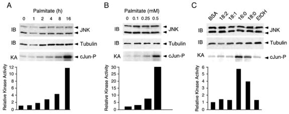
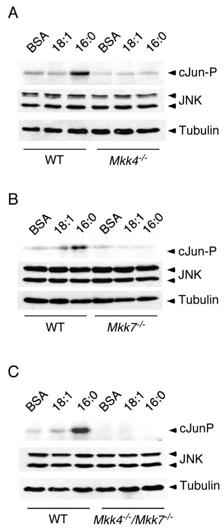
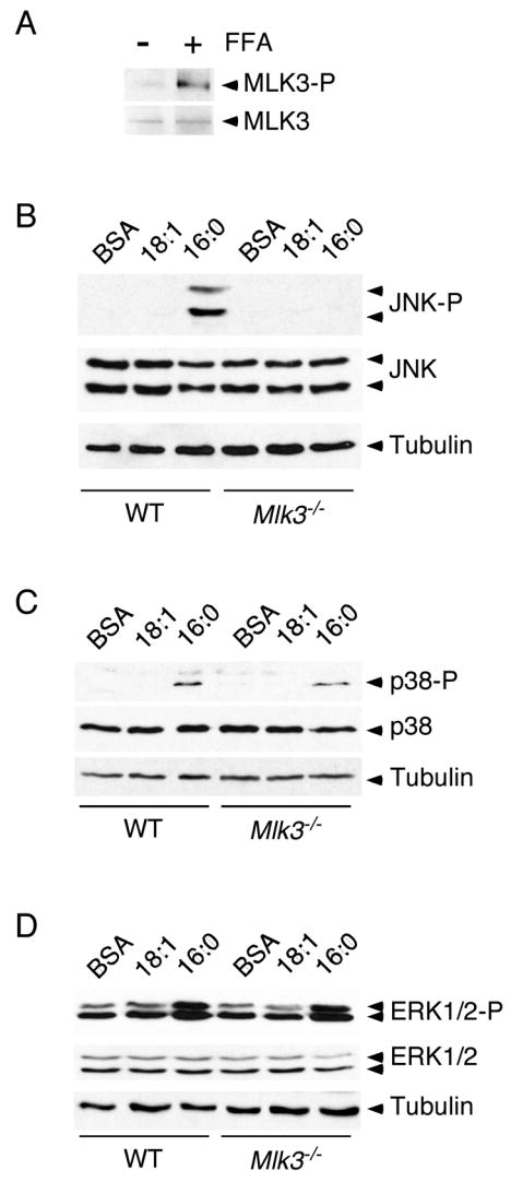
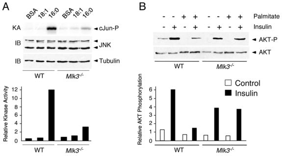
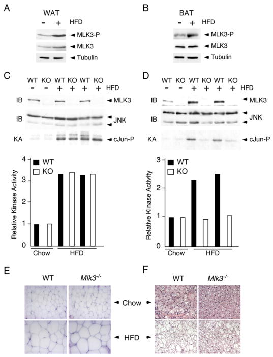
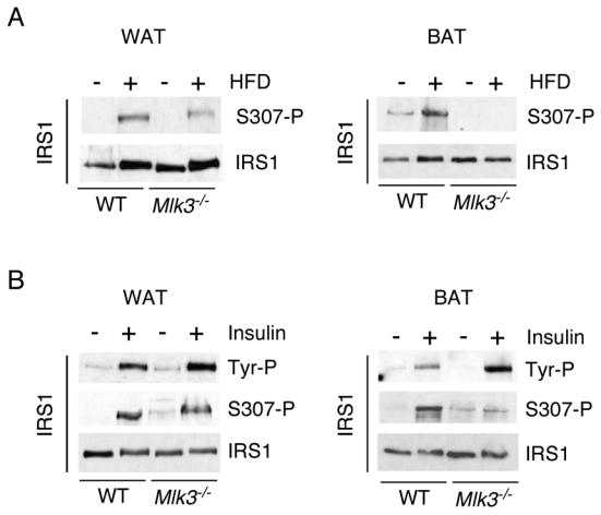
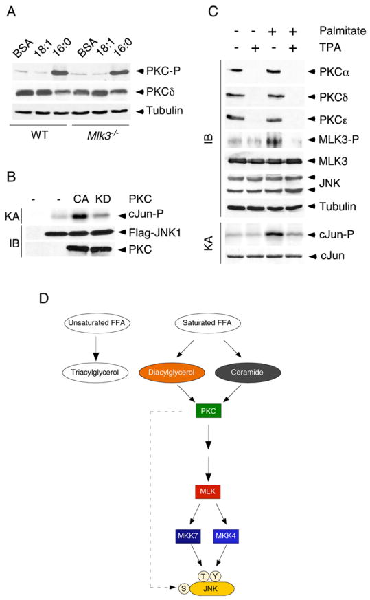
Saturated free fatty acid (FFA) is a major source of metabolic stress that activates the c-Jun NH(2)-terminal kinase (JNK). This FFA-stimulated JNK pathway is relevant to hallmarks of metabolic syndrome, including insulin resistance. Here we used gene ablation studies in mice to demonstrate a central role for mixed-lineage protein kinases (MLK) in this signaling pathway. Saturated FFA causes protein kinase C (PKC)-dependent activation of MLK3 that subsequently causes increased JNK activity by a mechanism that requires the MAP kinase kinases MKK4 and MKK7. Loss of PKC, MLK3, MKK4, or MKK7 expression prevents FFA-stimulated JNK activation. Together, these data establish a signaling pathway that mediates effects of metabolic stress on insulin resistance.


Figures

Figure 1
Figure 1. JNK is activated by saturated free fatty acids
A) MEF were treated with 0.5mM palmitate for the indicated times. The expression of JNK was examined by immunoblot analysis. JNK activity was measured in a kinase assay (KA) using [γ-32P]ATP and cJun as substrates. B) MEF were treated with different concentrations of palmitate for 16h. The expression of JNK was examined by immunoblot analysis. JNK activity was measured in a kinase assay (KA) using [γ-32P]ATP and cJun as substrates. C) MEF were treated with 0.5mM linoleate (18:2), oleate (18:1), palmitate (16:0) and stearic acid (18:0) for 16h. A solvent control (ethanol, Etoh) for the treatment with steric acid is shown. The expression of JNK was examined by immunoblot analysis. JNK activity was measured in a kinase assay (KA) using [γ-32P]ATP and cJun as substrates.
Figure 2
Figure 2. FFA-induced JNK activation is mediated by MKK4 and MKK7
WT or MKK-deficient MEF were treated with 0.5 mM oleic acid (18:1) or 0.5 mM palmitic acid (16:0) for 16 h. The expression of JNK was examined by immunoblot analysis. JNK activity was measured in an in vitro kinase assay (KA) using [γ-32P]ATP and cJun as substrates. A) WT and Mkk4−/− MEF. B) WT and Mkk7−/− MEF. C) WT and Mkk4−/− Mkk7−/− MEF.
Figure 3
Figure 3. JNK activation by saturated FFA is MLK3 dependent
(A) Wild-type MEF were treated (16 h) with 0.5 mM palmitic acid. The expression of MLK3 and phosphorylation of the MLK3 T-loop (Thr-277 and Ser281) was examined by immunoblot analysis. (B - D) Wild-type (WT) or Mlk3−/− MEF were treated (16 h) with 0.5 mM oleic acid (18:1) or 0.5 mM palmitic acid (16:0). The phosphorylation and expression of JNK (B), p38 MAPK (C), and ERK1/2 (D) was examined by immunoblot analysis.
Figure 4
Figure 4. MLK3-deficient cells are protected against FFA-induced insulin resistance
A) WT or Mlk3−/− MEF were treated with 0.5 mM oleic acid (18:1) or 0.5 mM palmitic acid (16:0) for 16 h. The expression of JNK was examined by immunoblot analysis. JNK activity was measured in a kinase assay (KA) using [γ-32P]ATP and cJun as substrates. B) WT or Mlk3−/− MEF were pretreated (16 h) with BSA or 0.5mM palmitate. After incubation with 100nM insulin for 30 min, the cells were harvested and AKT expression and phosphorylation at Ser-473 were examined by immunoblot analysis.
Figure 5
Figure 5. FFA causes MLK3 and JNK activation
A, B) Wild-type mice were maintained (16 weeks) on a standard diet or on a high fat diet (HFD). MLK3 expression, MLK3 T-loop phosphorylation (Thr-277 and Ser-281), and Tubulin expression in white (epididymal) fat (WAT) and brown (interscapular) fat (BAT) was examined by immunoblot analysis. C,D) White adipose tissue (C) and brown adipose tissue (D) of wild-type mice (WT) and Mlk3−/−mice (KO) maintained (16 weeks) on a standard diet (chow) and on a high fat diet (HFD) was examined by immunoblot analysis (IB) using antibodies to JNK and MLK3. JNK activity was measured in a kinase assay (KA) using [γ-32P]ATP and cJun as substrates. E,F) Representative histological sections of white adipose tissue (E) and brown adipose tissue (F) stained with hematoxylin and eosin from wild-type and Mlk3−/− mice fed a standard or high fat diet for 16 wk.
Figure 6
Figure 6. MLK3 is required for inhibitory phosphorylation of IRS1 on Ser 307
A) IRS1 expression and phosphorylation on Ser-307 in white epididymal adipose tissue (WAT) and brown interscapular adipose tissue (BAT) of wild-type and Mlk3−/− mice maintained (16 weeks) on a standard diet and on a high fat diet (HFD) was examined by immunoblot analysis. B) Wild-type and Mlk3−/− mice were fasted overnight and then treated (30 mins) with insulin (1.5 units/Kg). Extracts prepared from the fat pads (WAT and BAT) were examined by immunoblot analysis using antibodies to IRS1, tyrosine phosphorylated IRS1 (Tyr-P), and IRS1 phosphorylated on Ser-307.
Figure 7
Figure 7. PKC is required for MLK3 and JNK activation by FFA
A) Wild-type or Mlk3−/− MEF were treated (16 h) with BSA or with 0.5 mM oleic acid (18:1) or 0.5 mM palmitic acid (16:0). The phosphorylation and expression of PKCδ was examined by immunoblot analysis. B) Flag-tagged JNK1 and constitutively active or kinase-inactive PKCε were co-expressed in HEK 293 cells. The expression of JNK1 and PKC was examined by immunoblot analysis. JNK activity was measured in an IP-kinase assay (KA) using [γ-32P]ATP and cJun as substrates. C) PKCζ−/− MEF were pre-treated without and with 1 μM TPA (24 h) and then treated without and with 0.5 mM palmitate (16 h). The expression of PKCα, PKCδ, PKCε, MLK3, phospho-MLK3, JNK, phospho-JNK, and Tubulin was examined by immunoblot analysis. JNK activity was measured in a kinase assay (KA) using [γ-32P]ATP and cJun as substrates. D) Schematic illustration of a JNK signaling pathway that is activated by saturated FFA and is mediated by PKC, MLK, and MKK4/7.

References

    1. Aguirre V, Uchida T, Yenush L, Davis R, White MF. The c-Jun NH(2)-terminal kinase promotes insulin resistance during association with insulin receptor substrate-1 and phosphorylation of Ser(307) J Biol Chem. 2000;275:9047–9054. - PubMed
    1. Aguirre V, Werner ED, Giraud J, Lee YH, Shoelson SE, White MF. Phosphorylation of Ser307 in insulin receptor substrate-1 blocks interactions with the insulin receptor and inhibits insulin action. J Biol Chem. 2002;277:1531–1537. - PubMed
    1. Arner P. Insulin resistance in type 2 diabetes: role of fatty acids. Diabetes Metab Res Rev. 2002;18(Suppl 2):S5–9. - PubMed
    1. Barthwal MK, Sathyanarayana P, Kundu CN, Rana B, Pradeep A, Sharma C, Woodgett JR, Rana A. Negative regulation of mixed lineage kinase 3 by protein kinase B/AKT leads to cell survival. J Biol Chem. 2003;278:3897–3902. - PubMed
    1. Boden G. Effects of free fatty acids (FFA) on glucose metabolism: significance for insulin resistance and type 2 diabetes. Exp Clin Endocrinol Diabetes. 2003;111:121–124. - PubMed

Publication types

MeSH terms