

Referenceless MR thermometry for monitoring thermal ablation in the prostate
- PMID: 17679332
- PMCID: PMC2780365
- DOI: 10.1109/TMI.2007.892647
Referenceless MR thermometry for monitoring thermal ablation in the prostate
Abstract
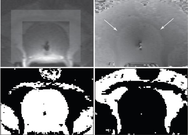
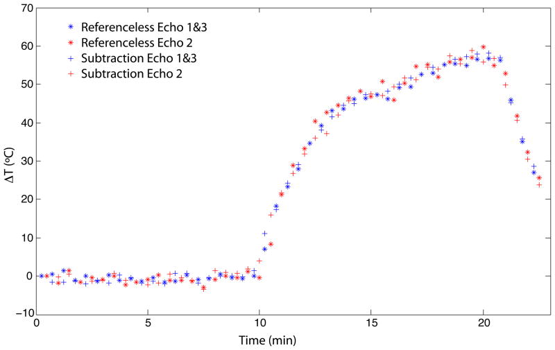
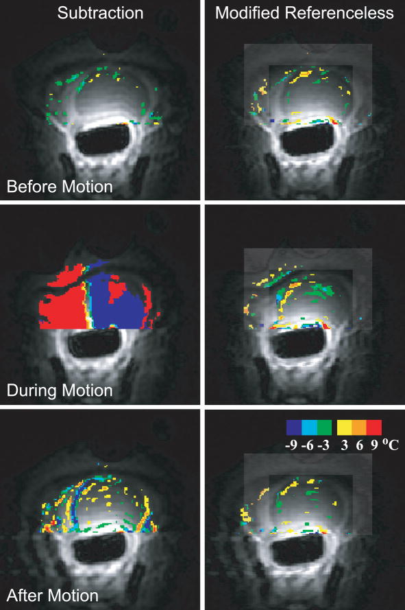
Referenceless proton resonance frequency (PRF) shift thermometry provides a means to measure temperature changes during minimally invasive thermotherapy that is inherently robust to motion and tissue displacement. However, if the referenceless method is used to determine temperature changes during prostate ablation, phase gaps between water and fat in image regions used to determine the background phase can confound the phase estimation. We demonstrate an extension to referenceless thermometry which eliminates this problem by allowing background phase estimation in the presence of phase discontinuities between aqueous and fatty tissue. In this method, images are acquired with a multiecho sequence and binary water and fat maps are generated from a Dixon reconstruction. For the background phase estimation, water and fat regions are treated separately and the phase offset between the two tissue types is determined. The method is demonstrated feasibile in phantoms and during in vivo thermal ablation of canine prostate.
Figures

Similar articles
-
Referenceless PRF shift thermometry.Magn Reson Med. 2004 Jun;51(6):1223-31. doi: 10.1002/mrm.20090. Magn Reson Med. 2004. PMID: 15170843
-
Reweighted ℓ1 referenceless PRF shift thermometry.Magn Reson Med. 2010 Oct;64(4):1068-77. doi: 10.1002/mrm.22502. Magn Reson Med. 2010. PMID: 20564600 Free PMC article.
-
Hybrid referenceless and multibaseline subtraction MR thermometry for monitoring thermal therapies in moving organs.Med Phys. 2010 Sep;37(9):5014-26. doi: 10.1118/1.3475943. Med Phys. 2010. PMID: 20964221 Free PMC article.
-
Technical advances in motion-robust MR thermometry.Magn Reson Med. 2024 Jul;92(1):15-27. doi: 10.1002/mrm.30057. Epub 2024 Mar 19. Magn Reson Med. 2024. PMID: 38501903 Free PMC article. Review.
-
Thermal monitoring: invasive, minimal-invasive and non-invasive approaches.Int J Hyperthermia. 2006 May;22(3):255-62. doi: 10.1080/02656730600661149. Int J Hyperthermia. 2006. PMID: 16754347 Review.
Cited by
-
Correction of breathing-induced errors in magnetic resonance thermometry of hyperthermia using multiecho field fitting techniques.Med Phys. 2010 Dec;37(12):6300-9. doi: 10.1118/1.3515462. Med Phys. 2010. PMID: 21302786 Free PMC article.
-
Percutaneous MR-guided prostate cancer cryoablation technical updates and literature review.BJR Open. 2019 Jun 3;1(1):20180043. doi: 10.1259/bjro.20180043. eCollection 2019. BJR Open. 2019. PMID: 33178928 Free PMC article. Review.
-
An anatomically realistic temperature phantom for radiofrequency heating measurements.Magn Reson Med. 2015 Jan;73(1):442-50. doi: 10.1002/mrm.25123. Epub 2014 Feb 18. Magn Reson Med. 2015. PMID: 24549755 Free PMC article.
-
Spatiotemporal filtering of MR-temperature artifacts arising from bowel motion during transurethral MR-HIFU.Med Phys. 2014 Nov;41(11):113302. doi: 10.1118/1.4897382. Med Phys. 2014. PMID: 25370670 Free PMC article.
-
EPI proton resonant frequency temperature mapping at 0.5T in the brain: Comparison to single-echo gradient recalled echo.Magn Reson Med. 2025 Apr;93(4):1733-1740. doi: 10.1002/mrm.30373. Epub 2024 Nov 11. Magn Reson Med. 2025. PMID: 39529375 Free PMC article.
References
-
- Beerlage HP, Thuroff S, Madersbacher S, Zlotta AR, Aus G, de Reijke TM, de la Rosette JJ. Current status of minimally invasive treatment options for localized prostate carcinoma. Eur Urol. 2000;37(1):2–13. - PubMed
-
- Shinohara K. Thermal ablation of prostate diseases: advantages and limitations. Int J Hyperthermia. 2004 Nov;20(7):679–97. - PubMed
-
- Larson BT, Bostwick DG, Corica AG, Larson TR. Histological changes of minimally invasive procedures for the treatment of benign prostatic hyperplasia and prostate cancer: clinical implications. J Urol. 2003 Jul;170(1):12–9. - PubMed
-
- Ishihara Y, Calderon A, Watanabe H, Okamoto K, Suzuki Y, Kuroda K, Suzuki Y. A precise and fast temperature mapping using water proton chemical shift. Magn Reson Med. 1995;34(6):814–23. - PubMed
-
- De Poorter J, De Wagter C, De Deene Y, Thomsen C, Stahlberg F, Achten E. Noninvasive MRI thermometry with the proton resonance frequency (PRF) method: in vivo results in human muscle. Magn Reson Med. 1995;33(1):74–81. - PubMed
Publication types
MeSH terms
Grants and funding
LinkOut - more resources
Full Text Sources
Other Literature Sources
Medical
