

Development of distinct control networks through segregation and integration
- PMID: 17679691
- PMCID: PMC1940033
- DOI: 10.1073/pnas.0705843104
Development of distinct control networks through segregation and integration
Abstract
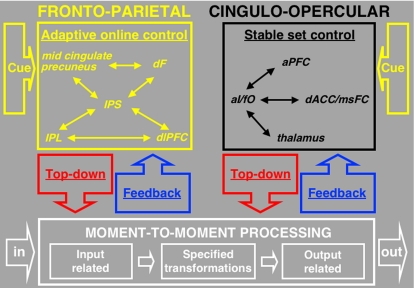
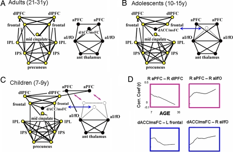
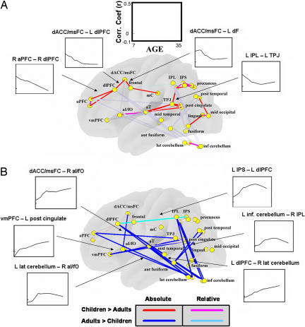
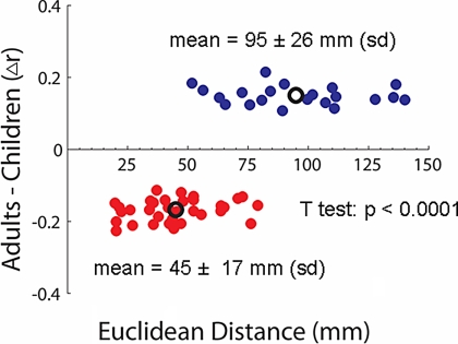
Human attentional control is unrivaled. We recently proposed that adults depend on distinct frontoparietal and cinguloopercular networks for adaptive online task control versus more stable set control, respectively. During development, both experience-dependent evoked activity and spontaneous waves of synchronized cortical activity are thought to support the formation and maintenance of neural networks. Such mechanisms may encourage tighter "integration" of some regions into networks over time while "segregating" other sets of regions into separate networks. Here we use resting state functional connectivity MRI, which measures correlations in spontaneous blood oxygenation level-dependent signal fluctuations between brain regions to compare previously identified control networks between children and adults. We find that development of the proposed adult control networks involves both segregation (i.e., decreased short-range connections) and integration (i.e., increased long-range connections) of the brain regions that comprise them. Delay/disruption in the developmental processes of segregation and integration may play a role in disorders of control, such as autism, attention deficit hyperactivity disorder, and Tourette's syndrome.
Conflict of interest statement
The authors declare no conflict of interest.
Figures
Similar articles
-
Functional Connectivity of Frontoparietal and Salience/Ventral Attention Networks Have Independent Associations With Co-occurring Attention-Deficit/Hyperactivity Disorder Symptoms in Children With Autism.Biol Psychiatry Cogn Neurosci Neuroimaging. 2019 Apr;4(4):343-351. doi: 10.1016/j.bpsc.2018.12.012. Epub 2019 Jan 9. Biol Psychiatry Cogn Neurosci Neuroimaging. 2019. PMID: 30777604 Free PMC article.
-
Developmental differences in higher-order resting-state networks in Autism Spectrum Disorder.Neuroimage Clin. 2014 May 14;4:820-7. doi: 10.1016/j.nicl.2014.05.007. eCollection 2014. Neuroimage Clin. 2014. PMID: 24936432 Free PMC article.
-
Integration and segregation across large-scale intrinsic brain networks as a marker of sustained attention and task-unrelated thought.Neuroimage. 2021 Apr 1;229:117610. doi: 10.1016/j.neuroimage.2020.117610. Epub 2021 Jan 6. Neuroimage. 2021. PMID: 33418073
-
Brain connectivity and visual attention.Brain Connect. 2013;3(4):317-38. doi: 10.1089/brain.2012.0139. Epub 2013 Jun 8. Brain Connect. 2013. PMID: 23597177 Free PMC article. Review.
-
Experience during adolescence shapes brain development: From synapses and networks to normal and pathological behavior.Neurotoxicol Teratol. 2019 Nov-Dec;76:106834. doi: 10.1016/j.ntt.2019.106834. Epub 2019 Sep 7. Neurotoxicol Teratol. 2019. PMID: 31505230 Review.
Cited by
-
Effects of preterm birth on intrinsic fluctuations in neonatal cerebral activity examined using optical imaging.PLoS One. 2013 Jun 28;8(6):e67432. doi: 10.1371/journal.pone.0067432. Print 2013. PLoS One. 2013. PMID: 23840698 Free PMC article.
-
Reorganization of Brain Networks in Aging and Age-related Diseases.Aging Dis. 2012 Apr;3(2):181-93. Epub 2011 Nov 28. Aging Dis. 2012. PMID: 22724079 Free PMC article.
-
The nuisance of nuisance regression: spectral misspecification in a common approach to resting-state fMRI preprocessing reintroduces noise and obscures functional connectivity.Neuroimage. 2013 Nov 15;82:208-25. doi: 10.1016/j.neuroimage.2013.05.116. Epub 2013 Jun 6. Neuroimage. 2013. PMID: 23747457 Free PMC article.
-
BrainNet Viewer: a network visualization tool for human brain connectomics.PLoS One. 2013 Jul 4;8(7):e68910. doi: 10.1371/journal.pone.0068910. Print 2013. PLoS One. 2013. PMID: 23861951 Free PMC article.
-
Impaired Bottom-Up Effective Connectivity Between Amygdala and Subgenual Anterior Cingulate Cortex in Unmedicated Adolescents with Major Depression: Results from a Dynamic Causal Modeling Analysis.Brain Connect. 2015 Dec;5(10):608-19. doi: 10.1089/brain.2014.0312. Epub 2015 Sep 28. Brain Connect. 2015. PMID: 26050933 Free PMC article.
References
Publication types
MeSH terms
Substances
Grants and funding
LinkOut - more resources
Full Text Sources
Other Literature Sources
