Review
. 2007 Sep;8(2-3):85-97.
doi: 10.1007/s10969-007-9024-5. Epub 2007 Aug 7.

Structural proteomics of the SARS coronavirus: a model response to emerging infectious diseases

Affiliations
Review

Structural proteomics of the SARS coronavirus: a model response to emerging infectious diseases

Mark Bartlam et al. J Struct Funct Genomics. 2007 Sep.

Abstract

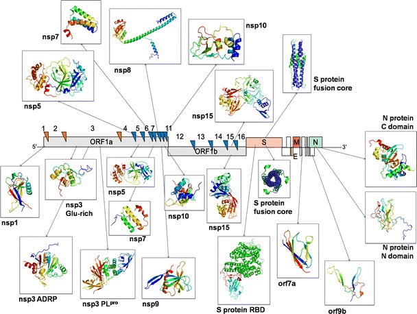
A number of structural genomics/proteomics initiatives are focused on bacterial or viral pathogens. In this article, we will review the progress of structural proteomics initiatives targeting the SARS coronavirus (SARS-CoV), the etiological agent of the 2003 worldwide epidemic that culminated in approximately 8,000 cases and 800 deaths. The SARS-CoV genome encodes 28 proteins in three distinct classes, many of them with unknown function and sharing low similarity to other proteins. The structures of 16 SARS-CoV proteins or functional domains have been determined to date. Remarkably, eight of these 16 proteins or functional domains have novel folds, indicating the uniqueness of the coronavirus proteins. The results of SARS-CoV structural proteomics initiatives will have several profound biological impacts, including elucidation of the structure-function relationships of coronavirus proteins; identification of targets for the design of anti-viral compounds against SARS-CoV and other coronaviruses; and addition of new protein folds to the fold space, with further understanding of the structure-function relationships for several new protein families. We discuss the use of structural proteomics in response to emerging infectious diseases such as SARS-CoV and to increase preparedness against future emerging coronaviruses.


Figures

Fig. 1
Fig. 1
Summary of SARS-CoV protein structures to date. The SARS-CoV genome is shown surrounded by the available structures of SARS-CoV proteins (drawn in ribbon representation): nsp1, nsp3 (Glu-rich, ADRP and PLpro domains), nsp5, nsp7, nsp8, nsp9, nsp10, nsp15, Spike protein (receptor binding domain and fusion core), N-protein (N-terminal RNA-binding domain and C-terminal dimerization domain), orf7a and orf9b. Orange and blue triangles represent PLpro (nsp3) and Mpro (nsp5) cleavage sites, respectively. Structures shown above the genome (nsp5, nsp7, nsp8, nsp10, nsp15, S-protein fusion core) were solved by Zihe Rao and colleagues in China. Representative structures shown below the genome were solved by other groups. Structures are not drawn to scale
Fig. 2
Fig. 2
Functional oligomers of SARS-CoV proteins. (A) Nsp5, the main protease (Mpro). SARS-CoV Mpro, shown in ribbon representation, is active as a dimer. (B) Nsp9, the ssRNA binding protein. SARS-CoV nsp9, shown in ribbon representation, functions as a dimer. (C) Nsp10, a zinc finger protein. SARS-CoV nsp10, shown in ribbon representation, can exist as a dodecamer in solution. The active form of nsp10 remains to be determined. Zinc ions are shown as grey spheres. (D) Nsp15, the endoribonuclease. Nsp15, shown in ribbon representation, is active as a hexamer. (E) The S-protein fusion core. The HR1 and HR2 peptides together form a six-helix bundle characteristic of class I viral fusion proteins. (F) The N-protein dimerization domain. The C-terminal domain of the N-protein functions as a dimer
Fig. 3
Fig. 3
SARS-CoV protein–protein complexes. (A) The structure of the nsp7–nsp8 supercomplex. The complex assembly is formed by eight copies of nsp7 and nsp8. Nsp8 exists simultaneously in two conformations, termed nsp8I and nsp8II. Nsp7, nsp8I and nsp8II are shown in ribbon representation (top) and colored blue, green and orange respectively. The complex (below) is assembled from two tetramers: T1, formed between nsp7 and nsp8I (center, left); and T2, formed between nsp7 and nsp8II (center, right). A surface representation showing the charge distribution is also shown (below right), with positive charge colored in blue and negative charge colored in red. The positive charge distributed around the central channel of the nsp7–nsp8 complex is favourable for the passage of RNA. (B) The SARS-CoV S-protein receptor binding domain complexed with the receptor ACE2. The complex structure is shown in ribbon representation with the ACE2 receptor colored in green, the S-protein receptor binding domain (RBD) colored in blue and the S-protein receptor binding motif (RBM) colored in red

Similar articles

Cited by

References

    1. Drosten C, Gunther S, Preiser W, van der Werf S, Brodt HR, Becker S, Rabenau H, Panning M, Kolesnikova L, Fouchier RA, et al. N Engl J Med. 2003;348:1967–1976. doi: 10.1056/NEJMoa030747. - DOI - PubMed
    1. Ksiazek TG, Erdman D, Goldsmith CS, Zaki SR, Peret T, Emery S, Tong S, Urbani C, Comer JA, Lim W, et al. N Engl J Med. 2003;348:1953–1966. doi: 10.1056/NEJMoa030781. - DOI - PubMed
    1. Kuiken T, Fouchier RA, Schutten M, Rimmelzwaan GF, van Amerongen G, van Riel D, Laman JD, de Jong T, van Doornum G, Lim W, et al. Lancet. 2003;362:263–270. doi: 10.1016/S0140-6736(03)13967-0. - DOI - PMC - PubMed
    1. Peiris JS, Lai ST, Poon LL, Guan Y, Yam LY, Lim W, Nicholls J, Yee WK, Yan WW, Cheung MT, et al. Lancet. 2003;361:1319–1325. doi: 10.1016/S0140-6736(03)13077-2. - DOI - PMC - PubMed
    1. Prentice E, McAuliffe J, Lu X, Subbarao K, Denison MR. J Virol. 2004;78:9977–9986. doi: 10.1128/JVI.78.18.9977-9986.2004. - DOI - PMC - PubMed

Publication types

MeSH terms

Substances