. 2007 Sep;27(6):757-70.
doi: 10.1007/s10571-007-9162-0. Epub 2007 Aug 7.

Halothane increases non-vesicular [(3)H]dopamine release from brain cortical slices

Affiliations

Halothane increases non-vesicular [(3)H]dopamine release from brain cortical slices

Paulo H C Diniz et al. Cell Mol Neurobiol. 2007 Sep.

Abstract

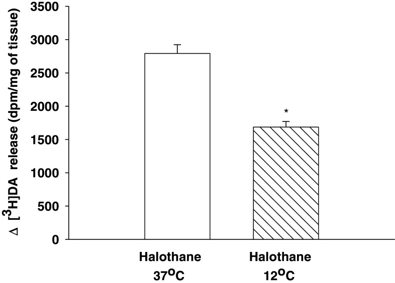
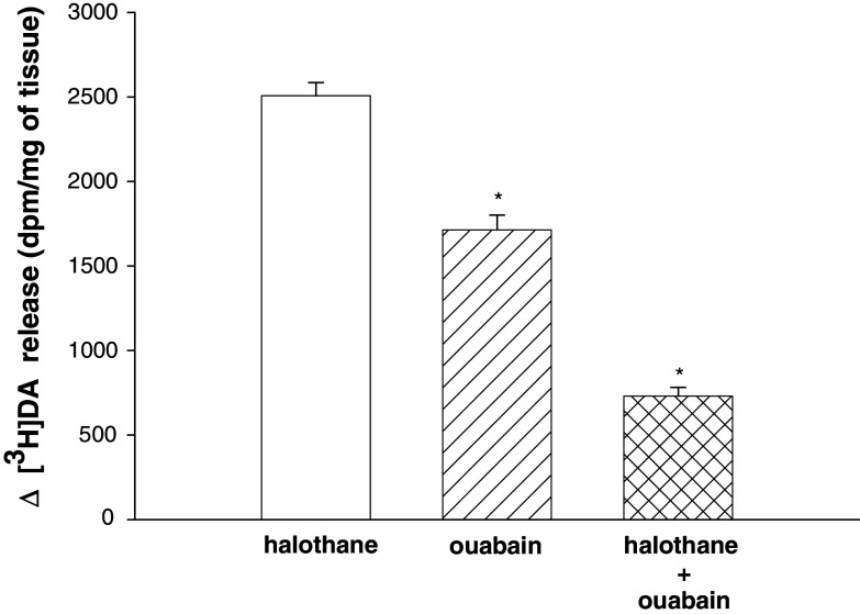
Experimental data suggest that halothane anesthesia is associated with significant changes in dopamine (DA) concentration in some brain regions but the mechanism of this effect is not well known. Rat brain cortical slices were labeled with [(3)H]DA to further characterize the effects of halothane on the release of this neurotransmitter from the central nervous system. Halothane induced an increase on the release of [(3)H]DA that was dependent on incubation time and anesthetic concentration (0.012, 0.024, 0.048, 0.072 and 0.096 mM). This effect was independent of extracellular or intracellular calcium. In addition, [(3)H]DA release evoked by halothane was not affected by TTX (blocker of voltage-dependent Na(+) channels) or reserpine (a blocker of vesicular monoamine transporter). These data suggest that [(3)H]DA release induced by halothane is non-vesicular and would be mediated by the dopamine transporter (DAT) and norepinephrine transporter (NET). GBR 12909 and nomifensine, inhibitors of DAT, decreased the release of [(3)H]DA evoked by halothane. Nisoxetine, a blocker of NET, reduced the release of [(3)H]DA induced by halothane. In addition, GBR 12909, nisoxetine and, halothane decrease the uptake of [(3)H]DA into rat brain cortical slices. A decrease on halothane-induced release of [(3)H]DA was also observed when the brain cortical slices were incubated at low temperature and low extracellular sodium, which are known to interfere with the carrier-mediated release of the neurotransmitter. Ouabain, a Na(+)/K(+) ATPase pump inhibitor, which induces DA release through reverse transport, decreased [(3)H]DA release induced by halothane. It is suggested that halothane increases [(3)H]DA release in brain cortical slices that is mediated by DAT and NET present in the plasma membrane.


Figures

Fig. 1
Fig. 1
Effect of halothane on [3H]DA release from rat brain cortical slices. (A) Brain cortical slices (40 mg) loaded with [3H]DA were incubated for 10 min in KHM in the absence (control) or in the presence of halothane at a concentration of 0.012, 0.024, 0.048, 0.072 and 0.096 mM. The EC50 value for neurotransmitter release evoked by halothane is 0.025 mM. The Δ values represent the difference between the release of DA in the presence of halothane and in the absence of halothane (control). Values are mean ± SEM from three individual experiments performed in duplicate. For other details, see text. (B) Slices loaded with [3H]DA were incubated for 2.5, 5, 10, 20 or 30 min in KHM in the absence (control) or in the presence of 0.048 mM halothane. The Δ values represent the difference between the release of DA in the presence of halothane and in the absence of halothane (control). Values are mean ± SEM from three individual experiments performed in duplicate. For other details, see text
Fig. 2
Fig. 2
[3H]DA release induced by halothane is calcium-independent. Brain cortical slices loaded with [3H]DA were pre-incubated with KHM in the absence or in the presence of EGTA (2.0 mM) or Cd2+ (100 μM) for 15 min or, for 30 min with BAPTA-AM (50 μM). They were then incubated with halothane (0.048 mM) for 10 min. The experiments with EGTA and BAPTA-AM were performed in a calcium free medium. The Δ values represent the difference between the release of DA in the presence of halothane and control (first bar), the difference between the release of DA in the presence of halothane plus EGTA and EGTA alone (second bar), the difference between the release of DA in the presence of halothane plus Cd2+ and Cd2+ alone (third bar), and the difference between the release of DA in the presence of halothane plus BAPTA-AM and BAPTA-AM alone (fourth bar). Values are mean ± SEM from five experiments performed in duplicate. For other details, see text
Fig. 3
Fig. 3
TTX does not interfere with halothane-evoked [3H]DA release. Brain cortical slices loaded with [3H]DA were pre-incubated for 15 min in KHM in the absence (control) or in the presence of TTX (1 μM) and then exposed to halothane (0.048 mM) for 10 min. The Δ values represent the difference between the release of DA in the presence of halothane and control (first bar) and, the difference between the release of DA in the presence of halothane plus TTX and TTX alone (second bar). Values are mean ± SEM from three individual experiments performed in duplicate
Fig. 4
Fig. 4
Effect of reserpine on [3H]DA release induced by halothane. Brain cortical slices loaded with [3H]DA were pre-incubated for 15 min in KHM in the absence (control) or in the presence of reserpine (0.03 μM). The slices were exposed to reserpine for additional 15 min in KHM and then stimulated with halothane (0.048 mM) for 10 min. The Δ values represent the difference between the release of DA in the presence of halothane and control (first bar) and, the difference between the release of DA in the presence of halothane plus reserpine and reserpine alone (second bar). The results are expressed as mean ± SEM from three individual experiments performed in duplicate
Fig. 5
Fig. 5
Effect of DAT and NET inhibitors on [3H]DA release induced by halothane. Brain cortical slices loaded with [3H]DA were pre-incubated for 20 min in KHM in the absence (control) or in the presence of nomifensine (1 μM) GBR 12909 (10 μM), or nisoxetine (10 μM). The slices were then incubated for 10 min in the absence (control) or in the presence of 0.048 mM halothane. The Δ values represent the difference between the release of DA in the presence of halothane and control (first bar), the difference between the release of DA in the presence of halothane plus nomifensine and nomifensine alone (second bar), the difference between the release of DA in the presence of halothane plus GBR 12909 and GBR 12909 alone (third bar) and, the difference between the release of DA in the presence of halothane plus nisoxetine and nisoxetine alone (fourth bar). The results are expressed as mean ± SEM from three individual experiments performed in duplicate. *Different from halothane alone (P < 0.05)
Fig. 6
Fig. 6
[3H]DA uptake in the presence of halothane, GBR 12909 and nisoxetine. Brain cortical slices were loaded with 0.20 μCi [3H]DA in the absence (control) or in the presence of GBR 12909 (10 μM), nisoxetine (10 μM), GBR 12909 plus nisoxetine or halothane (0.048 mM) for 30 min and then centrifuged at 4°C. After the treatment of the slices with trichloroacetic acid, the content of [3H]DA was measured for each experimental condition. The results are expressed as mean ± SEM from five individual experiments performed in duplicate. *Different from control alone (P < 0.05); **different from the GBR 12909 or nisoxetine value (P < 0.05)
Fig. 7
Fig. 7
Effect of low temperature on halothane-evoked [3H]DA release. Brain cortical slices loaded with [3H]DA were pre-incubated for 5 min in KHM at 37°C or 12°C and, subsequently incubated in the presence of halothane (0.048 mM) for 10 min. The Δ values represent the difference between the release of DA in the presence of halothane and control at 37°C (first bar) and, the difference between the release of DA in the presence of halothane and control at 12°C (second bar). The results are expressed as mean ± SEM from three individual experiments performed in duplicate. *Different from halothane at 37°C (P < 0.05)
Fig. 8
Fig. 8
Extracellular sodium levels interfere with [3H]DA release evoked by halothane. Brain cortical slices loaded with [3H]DA were pre-incubated for 5 min in KHM at NaCl 136 mM (control) or NaCl (40 mM) and, subsequently incubated in the presence of halothane (0.048 mM) for 10 min. The Δ values represent the difference between the release of DA in the presence of halothane and control with NaCl 136 mM (first bar) and, the difference between the release of DA in the presence of halothane and control with NaCl 40 mM (second bar). The results are expressed as mean ± SEM from three individual experiments performed in duplicate. *Different from halothane with NaCl 136 mM (P < 0.05)
Fig. 9
Fig. 9
Effect of ouabain on halothane-evoked [3H]DA release. Brain cortical slices loaded with [3H]DA were pre-incubated for 20 min in KHM in the absence (control) or in the presence of ouabain (100 μM). They were then stimulated with 0.048 mM halothane for additional 10 min. The Δ values represent the difference between the release of DA in the presence of halothane and control (first bar), the difference between the release of DA in the presence of ouabain and control (second bar) and, the difference between the release of DA in the presence of halothane plus ouabain and ouabain alone (third bar). The results are expressed as mean ± SEM from three individual experiments performed in duplicate. *Different from halothane alone (P < 0.05)

Similar articles

Cited by

References

    1. Adachi YU, Watanabe K, Satoh T, Vizi ES (2001a) Halothane potentiates the effect of methamphetamine and nomifensine on extracellular dopamine level in rat striatum. Br J Anaesth 86:837–845 - PubMed
    1. Adachi YU, Watanabe K, Higuchi H, Satoh T, Zsilla G (2001b) Halothane decreases impulse-dependent but not cytoplasmic release of dopamine from rat striatal slices. Brain Res Bull 56:521–524 - PubMed
    1. Adachi YU, Yamada S, Satomoto M, Higuchi H, Watanabe K, Kazama T (2005) Isoflurane anesthesia induces biphasic effect on dopamine release in the rat striatum. Brain Res Bull 67:176–181 - PubMed
    1. Adler EM, Augustine GJ, Duffy SN, Charlton MP (1991) Alien intracellular calcium chelators attenuate neurotransmitter release at the squid giant synapse. J Neurosci 11:1496–1507 - PMC - PubMed
    1. Andersen PH (1989) The dopamine inhibitor GBR 12909. Selectivity and molecular mechanism of action. Eur J Pharmacol 166:493–504 - PubMed

Publication types

MeSH terms

LinkOut - more resources