Comparative Study
. 2007 Nov 1;407(3):435-50.
doi: 10.1042/BJ20070579.

Differential modulation of Alzheimer's disease amyloid beta-peptide accumulation by diverse classes of metal ligands

Affiliations
Comparative Study

Differential modulation of Alzheimer's disease amyloid beta-peptide accumulation by diverse classes of metal ligands

Aphrodite Caragounis et al. Biochem J. .

Abstract

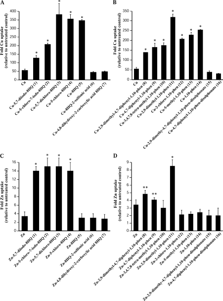
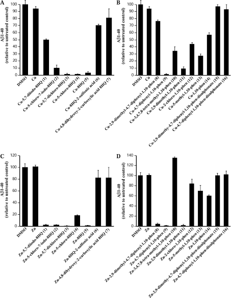
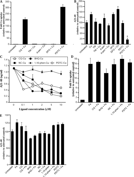
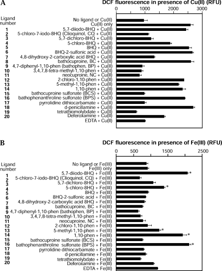
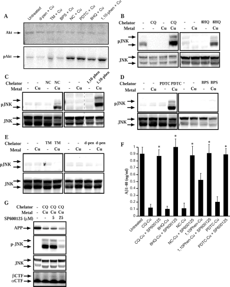
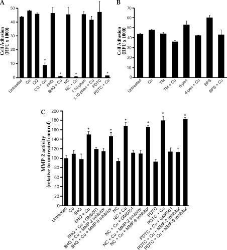
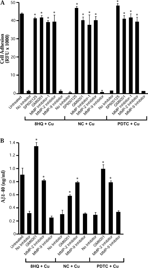
Biometals have an important role in AD (Alzheimer's disease) and metal ligands have been investigated as potential therapeutic agents for treatment of AD. In recent studies the 8HQ (8-hydroxyquinoline) derivative CQ (clioquinol) has shown promising results in animal models and small clinical trials; however, the actual mode of action in vivo is still being investigated. We previously reported that CQ-metal complexes up-regulated MMP (matrix metalloprotease) activity in vitro by activating PI3K (phosphoinositide 3-kinase) and JNK (c-jun N-terminal kinase), and that the increased MMP activity resulted in enhanced degradation of secreted Abeta (amyloid beta) peptide. In the present study, we have further investigated the biochemical mechanisms by which metal ligands affect Abeta metabolism. To achieve this, we measured the effects of diverse metal ligands on cellular metal uptake and secreted Abeta levels in cell culture. We report that different classes of metal ligands including 8HQ and phenanthroline derivatives and the sulfur compound PDTC (pyrrolidine dithiocarbamate) elevated cellular metal levels (copper and zinc), and resulted in substantial loss of secreted Abeta. Generally, the ability to inhibit Abeta levels correlated with a higher lipid solubility of the ligands and their capacity to increase metal uptake. However, we also identified several ligands that potently inhibited Abeta levels while only inducing minimal change to cellular metal levels. Metal ligands that inhibited Abeta levels [e.g. CQ, 8HQ, NC (neocuproine), 1,10-phenanthroline and PDTC] induced metal-dependent activation of PI3K and JNK, resulting in JNK-mediated up-regulation of metalloprotease activity and subsequent loss of secreted Abeta. The findings in the present study show that diverse metal ligands with high lipid solubility can elevate cellular metal levels resulting in metalloprotease-dependent inhibition of Abeta. Given that a structurally diverse array of ligands was assessed, the results are consistent with the effects being due to metal transport rather than the chelating ligand interacting directly with a receptor.


Figures

Figure 1
Figure 1. Effect of metal ligands on Cu and Zn uptake into APP-CHO cells
Cells were treated with 25 μM ligand–metal complexes for 6 h and metal levels were determined in cell pellets by ICP-MS. Metal uptake is given as the fold increase compared with vehicle-treated control. (A) Treatment of cells with 8HQ derivatives and Cu. (B) Treatment of cells with phenanthroline derivatives and Cu. (C) Treatment of cells with 8HQ derivatives and Zn. (D) Treatment of cells with phenanthroline derivatives and Zn. *P<0.01 and **P<0.05. phen, phenanthroline.
Figure 2
Figure 2. Effect of metal ligands on Aβ-(1–40) levels in APP-CHO cells
Cells were treated with 25 μM ligand–metal complexes for 6 h and Aβ-(1–40) levels were determined by ICP-MS in conditioned medium. (A) Treatment of cells with 8HQ derivatives and Cu. (B) Treatment of cells with phenanthroline derivatives and Cu. (C) Treatment of cells with 8HQ derivatives and Zn. (D) Treatment of cells with phenanthroline derivatives and Zn. phen, phenanthroline.
Figure 3
Figure 3. Effect of alternative ligand–metal complexes on metal uptake and Aβ-(1–40) levels in APP-CHO cells
Cells were treated with metal ligands with or without 25 μM metal for 6 h and metal uptake was determined by ICP-MS. Aβ-(1–40) levels were determined in conditioned medium by ELISA. (A) Cu uptake in cells after treatment with sulfur-containing or alternative metal ligands together with Cu. (B) Aβ-(1–40) levels in medium from cultures treated with TM, D-penicillsamine, DFO or PDTC. (C) Aβ-(1–40) levels in cells treated with low concentrations of CQ, 8HQ, NC, 1,10-phenanthroline or PDTC plus Cu. (D) Fe levels in cells treated with CQ, 8HQ, NC, 1,10-phenanthroline or PDTC and Fe(II). (E) Aβ-(1–40) levels in medium of cells treated with CQ, 8HQ, NC, 1,10-phenanthroline or PDTC and Fe(II). *P<0.01. d-pen, D-penicillamine; phen, phenanthroline.
Figure 4
Figure 4. DCF fluorescence measurement of hydrogen peroxide generation by metal–ligand complexes
(A) Ligands were incubated with Cu(II) and hydrogen peroxide generation (redox activity) was determined by DCF fluorescence. All samples contained ascorbate and HRP. Ligand and/or metal were added as indicated. No ligand induced higher levels of hydrogen peroxide than Cu(II) alone indicating a lack of redox activity towards Cu(II). (B) Ligands were incubated with Fe(III) and hydrogen peroxide generation was determined by DCF fluorescence. All samples contained ascorbate and HRP. Ligand and/or metal were added as indicated. Several ligands induced a significant increase in hydrogen peroxide compared with Fe(III) alone (*P<0.001). phen, phenanthroline.
Figure 5
Figure 5. Effect of CQ and metals on APP metabolism
(A) APP-CHO cells were treated with CQ and Cu, Zn or Fe(II) for 6 h. CQ alone or CQ and Cu or Zn induced a decrease in cellular APP expression. Co-treatment with Fe(II) inhibited the loss of APP expression induced by CQ. (B) Treatment of APP-CHO cells with CQ or CQ plus Cu induced a reduction in secreted APP (sAPP) levels in conditioned medium after 6 h. The loss of sAPP corresponded to the loss of cellular APP. Treatment with CQ, Cu or CQ and Cu had no effect on expression of APP C-terminal fragments (CTFs).
Figure 6
Figure 6. Activation of PI3K and JNK by diverse ligand–metal complexes in APP-CHO cells
Cells were treated with ligands plus Cu for 6 h and activation (phosphorylation) of JNK was determined by Western blot analysis in cell lysates. (A) Activation of PI3K (phospho-Akt formation) by Cu and 8HQ, NC, PDTC and 1,10-phenanthroline. Some activation was observed with TM plus Cu but not with BPS or D-penicillamine plus Cu. (B) Activation of JNK by CQ or 8HQ and Cu. (C) JNK activation by NC or 1,10-phenanthroline and Cu. (D) Activation of JNK by PDTC and Cu but not by BPS and Cu. (E) No activation of JNK by TM or D-penicillamine and Cu. (F) Treatment of cells with an inhibitor of JNK activation (SP600125; 25 μM) prevented the loss of Aβ-(1–40) induced by CQ, 8HQ, NC, 1,10-phenanthroline or PDTC and Cu. *P<0.01. (G) Treatment of cells with the JNK inhibitor, SP600125 inhibited JNK activation but did not restore the loss of APP expression induced by CQ–Cu complexes. The inhibitor also did not alter C-terminal APP processing. CTF, C-terminal fragment; d-pen, D-penicillamine; phen, phenanthroline.
Figure 7
Figure 7. Effect of metal ligands on cell adhesion and MMP activity in APP-CHO cells
Cells were treated with metal ligands and Cu for 6 h and cell adhesion was determined on a collagen type IV matrix. MMP-2 activity was measured by fluorometric assay in cell lysates. (A) Cell adhesion after treatment with CQ, 8HQ, NC, 1,10-phenanthroline or PDTC with or without Cu. (B) Cell adhesion after treatment with TM, D-penicillamine or BPS with or without Cu. (C) MMP-2 activity in cells after treatment with 8HQ, NC or PDTC and Cu. In each treatment, the broad-spectrum metalloprotease inhibitor GM6001 or the MMP-2 inhibitor prevented MMP activation (*P<0.01). d-pen, D-penicillamine; phen, phenanthroline.
Figure 8
Figure 8. Effect of JNK and MMP inhibitors on loss of cell adhesion and secreted Aβ levels in APP-CHO cells
Cells were treated with 8HQ, NC or PDTC and Cu for 6 h in the presence or absence of 25 μM SP600125 (JNK inhibitor), 20 μM GM6001 (broad-spectrum MMP inhibitor), 25 μM MMP inhibitor-I (broad-spectrum MMP inhibitor), 10 μM MMP-2 inhibitor-I or 10 μM MMP-9 inhibitor-I. (A) Inhibition of JNK or MMPs blocked the loss of cell adhesion induced by each metal ligand complexed with Cu (*P<0.01). (B) Inhibition of MMPs prevented the loss of secreted Aβ-(1–40) induced by each metal ligand complexed with Cu (*P<0.01).

References

    1. Bush A. I., Pettingell W. H., Multhaup G., d Paradis M., Vonsattel J. P., Gusella J. F., Beyreuther K., Masters C. L., Tanzi R. E. Rapid induction of Alzheimer A β amyloid formation by zinc. Science. 1994;265:1464–1467. - PubMed
    1. Glenner G. G., Wong C. W. Alzheimer's disease: initial report of the purification and characterization of a novel cerebrovascular amyloid protein. Biochem. Biophys. Res. Commun. 1984;120:885–890. - PubMed
    1. Masters C. L., Simms G., Weinman N. A., Multhaup G., McDonald B. L., Beyreuther K. Amyloid plaque core protein in Alzheimer disease and Down syndrome. Proc. Natl. Acad. Sci. U.S.A. 1985;82:4245–4249. - PMC - PubMed
    1. Atwood C. S., Scarpa R. C., Huang X., Moir R. D., Jones W. D., Fairlie D. P., Tanzi R. E., Bush A. I. Characterization of copper interactions with Alzheimer amyloid β peptides: identification of an attomolar-affinity copper binding site on amyloid β1–42. J. Neurochem. 2000;75:1219–1233. - PubMed
    1. Bush A. I., Multhaup G., Moir R. D., Williamson T. G., Small D. H., Rumble B., Pollwein P., Beyreuther K., Masters C. L. A novel zinc(II) binding site modulates the function of the βA4 amyloid protein precursor of Alzheimer's disease. J. Biol. Chem. 1993;268:16109–16112. - PubMed

Publication types