. 2007 Sep 1;406(2):265-71.
doi: 10.1042/BJ20070441.

TFIIB/SUA7(E202G) is an allele-specific suppressor of TBP1(E186D)

Affiliations

TFIIB/SUA7(E202G) is an allele-specific suppressor of TBP1(E186D)

Boon Shang Chew et al. Biochem J. .

Abstract

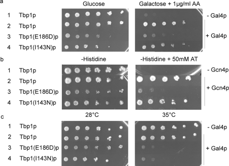
The TBP (TATA-box-binding protein), Tbp1p, plays a vital role in all three classes of transcription by RNA polymerases I-III. A TBP1(E186D) mutation had been described that affected interaction of Tbp1p with TFIIB (transcription factor IIB) and that caused slow-growth, temperature-sensitivity, 3-aminotriazole-sensitivity as well as a gal(-) phenotype. We used the TBP1(E186D) mutant for suppressor screens, and we isolated TFIIB/SUA7(E202G) as an allele-specific suppressor of all phenotypes caused by the TBP1(E186D) mutation. Our results show that the SUA7(E202G) mutation restored binding of TFIIB to Tbp1(E186D)p. In addition, we observed that Tbp1(E186D)p was expressed at a lower level than wild-type Tbp1p, and that SUA7(E202G) restored the protein level of Tbp1(E186D)p. This suggested that the TBP1(E186D) mutation might have generated its phenotypes by making Tbp1p the limiting factor for activated transcription. DNA microarray analysis indicated that the TBP1(E186D) temperature-sensitivity and slow-growth phenotypes might have been caused by insufficient amounts of Tbp1p for efficient transcription of the rRNA genes by RNA polymerase I.


Figures

Figure 1
Figure 1. The TBP1(E186D) mutation affects activation by Gal4p and Gcn4p and causes temperature-sensitivity
(a) Ten-fold serial dilutions of yeast cells expressing the depicted proteins were titrated on glucose and galactose plates containing 1 μg/ml antimycin A (AA) and incubated on the indicated medium at 28 °C for 10 days. (b) Ten-fold serial dilutions of yeast cells expressing the depicted proteins were titrated on the indicated medium and incubated at 28 °C for 10 days. (c) Ten-fold serial dilutions of yeast cells expressing the depicted proteins were titrated on glucose medium and incubated at 28 and 35 °C for 10 days.
Figure 2
Figure 2. SUA7(E202G) is an allele-specific suppressor of TBP1(E186D)
Ten-fold serial dilutions of yeast cells expressing the depicted proteins were dropped on to the indicated medium and incubated at the respective temperatures for 10 days.
Figure 3
Figure 3. The SUA7(E202G) mutation restores the protein interaction with TBP1(E186D) in vivo
Ten-fold serial dilutions of BY4741ΔW cells expressing the depicted proteins were titrated on the indicated plates and incubated at 39 °C for 3 days. Yeast cells expressing Tbp1(E186D)-Cub-RUra3p were incubated on a uracil-depleted plate containing 100 μM CuSO4 so that the growth of cells expressing Nub and Tbp1(E186D)-Cub-RUra3p was comparable with yeast cells expressing Nub and Tbp1-Cub-RUra3p.
Figure 4
Figure 4. The SUA7(E202G) mutation restores the protein levels of TBP1(E186D)
(a) Complementation of GST-fusion proteins in NLY2 strain. A yeast strain carrying a chromosomal deletion of TBP1 and expressing TBP1 from a URA3-marked single-copy vector was transformed with plasmids expressing the depicted proteins. Ten-fold serial dilutions of cells were titrated on glucose medium with and without FOA. The plates were incubated at 28 °C for 7 days. Growth on FOA indicated that the respective GST–Tbp1p fusion was able to complement the chromosomal TBP1 deletion. (b) GST pull-down and Western blot for the analysis of the in vitro protein–protein interaction between Sua7(E202G)p and Tbp1(E186D)p. Yeast cells expressing the depicted proteins were grown in a medium lacking tryptophan and leucine, and GST pull-down assays were performed with cell extracts followed by Western-blot analysis with anti-GST and anti-HA antibodies. (c) Western blot for the analysis of the expression of endogenous Tbp1p and Tbp1(E186D)p. Yeast cells expressing the depicted proteins were grown in medium lacking tryptophan and leucine, and Western-blot analysis was performed with anti-Tbp1p antibodies. As a loading control, all proteins were visualized with Coomassie Brilliant Blue.

Similar articles

Cited by

References

    1. Naar A. M., Lemon B. D., Tjian R. Transcriptional coactivator complexes. Annu. Rev. Biochem. 2001;70:475–501. - PubMed
    1. Woychik N. A., Hampsey M. The RNA polymerase II machinery: structure illuminates function. Cell. 2002;108:453–463. - PubMed
    1. Szutorisz H., Dillon N., Tora L. The role of enhancers as centres for general transcription factor recruitment. Trends Biochem. Sci. 2005;30:593–599. - PubMed
    1. Ptashne M. Regulation of transcription: from lambda to eukaryotes. Trends Biochem. Sci. 2006;30:275–279. - PubMed
    1. Santangelo G. M. Glucose signaling in Saccharomyces cerevisiae. Microbiol. Mol. Biol. Rev. 2006;70:253–282. - PMC - PubMed

Publication types

MeSH terms