Review
. 2008 Mar;7(2):129-51.
doi: 10.1111/j.1601-183X.2007.00342.x. Epub 2007 Aug 3.

Genetic findings in Parkinson's disease and translation into treatment: a leading role for mitochondria?

Affiliations
Review

Genetic findings in Parkinson's disease and translation into treatment: a leading role for mitochondria?

V Bogaerts et al. Genes Brain Behav. 2008 Mar.

Abstract

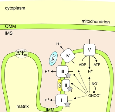
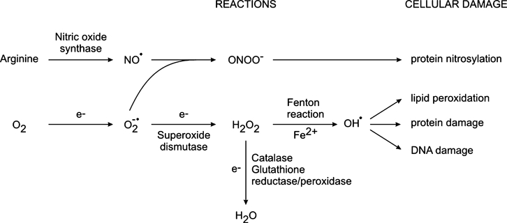
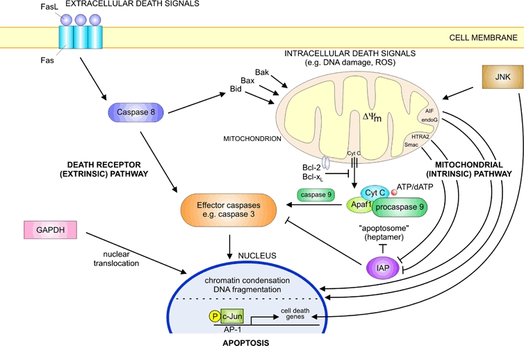
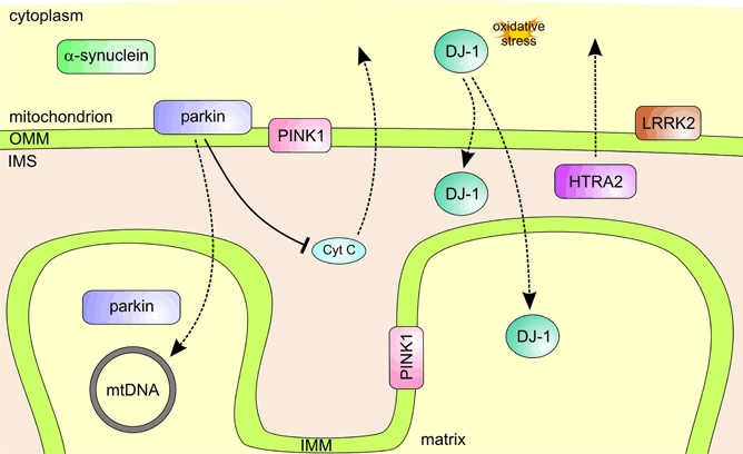
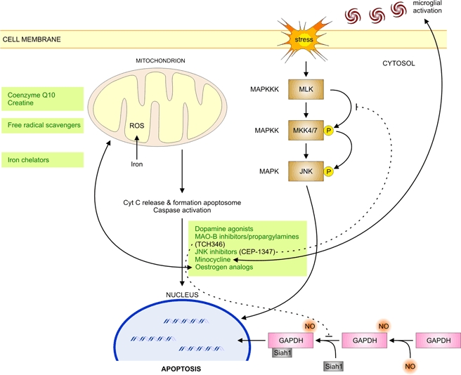
Parkinson's disease (PD) is a progressive neurodegenerative movement disorder and in most patients its aetiology remains unknown. Molecular genetic studies in familial forms of the disease identified key proteins involved in PD pathogenesis, and support a major role for mitochondrial dysfunction, which is also of significant importance to the common sporadic forms of PD. While current treatments temporarily alleviate symptoms, they do not halt disease progression. Drugs that target the underlying pathways to PD pathogenesis, including mitochondrial dysfunction, therefore hold great promise for neuroprotection in PD. Here we summarize how the proteins identified through genetic research (alpha-synuclein, parkin, PINK1, DJ-1, LRRK2 and HTRA2) fit into and add to our current understanding of the role of mitochondrial dysfunction in PD. We highlight how these genetic findings provided us with suitable animal models and critically review how the gained insights will contribute to better therapies for PD.


Figures

Figure 1
Figure 1
Mitochondrial structure and composition of the mitochondrial respiratory chain. The mitochondrial respiratory chain is a sequence of complexes found in the IMM that accepts electrons from electron donors such as NADH or succinate, shuttles these electrons across the IMM and creates a proton and electrochemical gradient. This gradient forms the basis of the inner mitochondrial transmembrane potential (ΔΨm) and is used to drive ATP synthesis by complex V of the respiratory chain. Complexes I, II and III generate ROS (indicated by grey stars). NO inhibits respiration by reversible binding to the oxygen binding site of complex IV (not shown) and is likely to be a physiological regulator of respiration. When cells are under oxidative stress, ROS will accumulate, react with NO, and form peroxynitrite (ONOO); a strong oxidant thought to be responsible for the ‘pathological actions’ of NO. ONOO inactivates the respiratory complexes (dotted lines), stimulates proton leakage through the IMM and might inhibit complex I by tyrosine nitration (for review see Brown & Borutaite 2002). Abbreviations: ΔΨm, inner mitochondrial transmembrane potential; ADP, adenosine 5′-diphosphate; CoQ, coenzyme Q; Cyt C, cytochrome c.
Figure 2
Figure 2
ROS. These include (1) free radicals (containing highly reactive unpaired electrons), such as superoxide (O2−·), nitric oxide (NO·) and hydroxyl radical (OH·); and (2) other molecular species, such as hydrogen peroxide (H2O2) and peroxynitrite (ONOO). O2−· is converted to H2O2, either spontaneously or through a reaction catalysed by SOD (Fridovich 1995). H2O2 may in turn be fully reduced to water, by catalase or glutathione reductase/peroxidase, or partially reduced to hydroxyl radicals (OH·). The latter reaction (Fenton reaction) occurs in the presence of reduced transition metals (e.g. Fe2+), which may again be re-reduced by O2−·, propagating the process (Liochev & Fridovich 1994). Alternatively O2−· can also react with NO radicals (NO·; produced by nitric oxide synthase during conversion of arginine to citrulline) to form peroxynitrite (ONOO) (Beckman & Koppenol 1996).
Figure 3
Figure 3
Schematic illustration of the major pathways leading to apoptosis. Apoptosis occurs through two main pathways. These are the death receptor (extrinsic) pathway which originates through the activation of cell-surface death receptors, for example Fas, and the mitochondrial (intrinsic) pathway which originates from mitochondrial release of cytochrome c. A distinct nuclear pathway of apoptosis arises through increased expression of GAPDH and its translocation from the cytoplasm to the nucleus, whereas the JNK signal-transduction pathway is activated in response to different stress stimuli. Bid, Bax and Bak represent pro-apoptotic Bcl-2 family members; Bcl-2 and Bcl-xL are antiapoptotic. Abbreviations: ΔΨm, inner mitochondrial transmembrane potential; AP-1, activating protein-1; Apaf1, apoptotic protease-activating factor 1; dATP, deoxyadenosine 5′-triphosphate; Cyt C, cytochrome c; endoG, endonuclease G; FasL, Fas ligand.
Figure 4
Figure 4
PD genes and their relation to mitochondria. Aggregation of mutant or overexpressed SNCA might be an upstream actor of mitochondrial alterations. Parkin associates with the OMM and was shown to play a role in mitochondrial biogenesis by regulating both transcription and replication of mtDNA. PINK1 has an N-terminal mitochondrial targeting motif and is localized to mitochondrial membranes, whereas oxidation of a key Cys-residue in DJ-1 leads to its relocalization to mitochondria. LRRK2 resides diffusely throughout the cytosol, but is partly associated with the OMM. HTRA2 resides in the IMS, wherefrom it is released upon apoptotic stimuli.
Figure 5
Figure 5
Entry points for PD therapy involving mitochondria. Green boxes highlight potential neuroprotective drugs at their respective action levels, and dotted lines indicate premature termination of clinical trials for promising neuroprotective drugs. Abbreviations: Cyt C, cytochrome c; MAPKK, MAPK kinase; MAPKKK, MAPKK kinase; MKK4/7, MAPK kinases 4/7; P, phosphorylated.

Similar articles

Cited by

References

    1. Abeliovich A, Schmitz Y, Farinas I, Choi-Lundberg D, Ho WH, Castillo PE, Shinsky N, Verdugo JMG, Armanini M, Ryan A, Hynes M, Phillips H, Sulzer D, Rosenthal A. Mice lacking alpha-synuclein display functional deficits in the nigrostriatal dopamine system. Neuron. 2000;25:239–252. - PubMed
    1. Abou-Sleiman PM, Healy DG, Quinn N, Lees AJ, Wood NW. The role of pathogenic DJ-1 mutations in Parkinson’s disease. Ann Neurol. 2003;54:283–286. - PubMed
    1. Abou-Sleiman PM, Healy DG, Wood NW. Causes of Parkinson’s disease: genetics of DJ-1. Cell Tissue Res. 2004;318:185–188. - PubMed
    1. Abou-Sleiman PM, Muqit MMK, McDonald NQ, Yang YX, Gandhi S, Healy DG, Harvey K, Harvey RJ, Deas E, Hatia K, Quinn N, Lees A, Latchman DS, Wood NW. A heterozygous effect for PINK1 mutations in Parkinson’s disease? Ann Neurol. 2006;60:414–419. - PubMed
    1. Akao Y, Maruyama W, Yi H, Shamoto-Nagai M, Youdim MBH, Naoi M. An anti-Parkinson’s disease drug, N-propargyl-1(R)-aminoindan (rasagiline), enhances expression of anti-apoptotic Bcl-2 in human dopaminergic SH-SY5Y cells. Neurosci Lett. 2002;326:105–108. - PubMed

Publication types