

Longitudinal study of keratoconus progression
- PMID: 17681291
- PMCID: PMC3597220
- DOI: 10.1016/j.exer.2007.06.016
Longitudinal study of keratoconus progression
Abstract
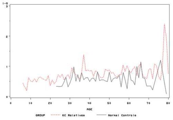
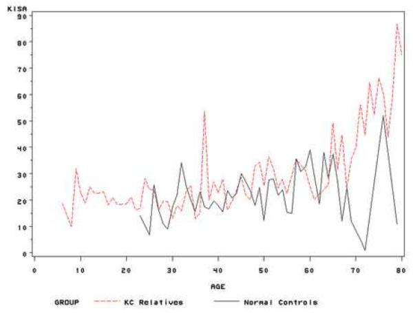
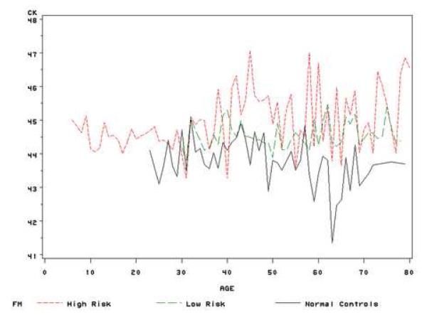
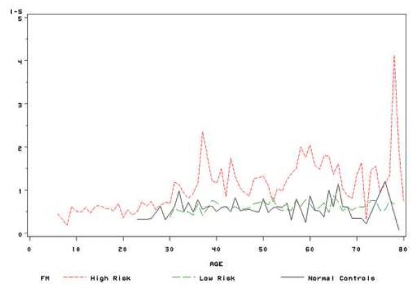
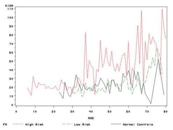
To determine if differences in topographic progression between unaffected keratoconus relatives and normal controls can predict factors associated with the development of keratoconus in a longitudinal study. We recruited 369 unaffected keratoconus relatives and 119 normal controls in Los Angeles. Both eyes of subjects were examined at baseline clinically and by quantitative videokeratography and at a period ranging from 1 year to 8 years. Progression to keratoconus was evaluated by quantitative videokeratography variables. Unaffected relatives had higher Central K (CK), I-S and KISA values and were younger than normal controls (CK: 44.70 vs 44.01, P<0.01; I-S: 0.76 vs 0.58, P<0.01, KISA: 29.97 vs 23.89, P=0.02; age: 34.8 vs 41.0, P<0.01) at baseline. All three indices significantly increased with age, and CK and KISA values were associated with a positive family history for keratoconus (P<0.001 for CK and P=0.05 for KISA), however, the two groups were not statistically different in progression of keratoconus. After grouping unaffected relatives as high risk (age< or = 30 or Central K > or = 47.2 and I-S> or =1.2 or KISA> or = 60) and low risk (age>30 and Central K<47.2 and I-S<1.2 and KISA< 60), relatives in the high risk group had a greater increase in CK and I-S values than those in the low risk group (CK: P=0.009; I-S: P<0.001), which indicated that there were significantly different rates of progression between two groups. Unaffected relatives had higher videokeratography indices than normal controls, but overall they did not progress to keratoconus quicker than normal controls. However, relatives in the high risk group may have a greater risk of progression to keratoconus.
Figures

References
-
- Edwards M, McGhee CN, Dean S. The genetics of keratoconus. Clin.Exp. Ophthalmol. 2001;29:345–351. - PubMed
-
- Falls HF, Allen AW. Dominantly inherited keratoconus. Journal of Genetic Human. 1969;17:317–324. - PubMed
-
- Holland DR, Maeda N, Hannush SB, Riveroll LH, Green MT, Klyce SD, Wilson SE. Unilateral keratoconus: incidence and quantitative topographic analysis. Ophthalmology. 1997;104:1409–13. - PubMed
Publication types
MeSH terms
Grants and funding
LinkOut - more resources
Full Text Sources
Medical
Research Materials
