Review
. 2007 Oct 1;623(1-2):83-97.
doi: 10.1016/j.mrfmmm.2007.06.009. Epub 2007 Jul 3.

DNA topoisomerase II, genotoxicity, and cancer

Affiliations
Review

DNA topoisomerase II, genotoxicity, and cancer

A Kathleen McClendon et al. Mutat Res. .

Abstract

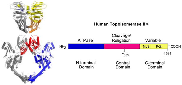
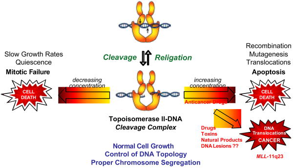
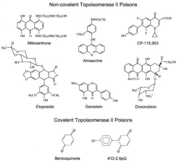
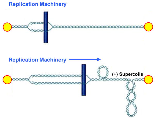
Type II topoisomerases are ubiquitous enzymes that play essential roles in a number of fundamental DNA processes. They regulate DNA under- and overwinding, and resolve knots and tangles in the genetic material by passing an intact double helix through a transient double-stranded break that they generate in a separate segment of DNA. Because type II topoisomerases generate DNA strand breaks as a requisite intermediate in their catalytic cycle, they have the potential to fragment the genome every time they function. Thus, while these enzymes are essential to the survival of proliferating cells, they also have significant genotoxic effects. This latter aspect of type II topoisomerase has been exploited for the development of several classes of anticancer drugs that are widely employed for the clinical treatment of human malignancies. However, considerable evidence indicates that these enzymes also trigger specific leukemic chromosomal translocations. In light of the impact, both positive and negative, of type II topoisomerases on human cells, it is important to understand how these enzymes function and how their actions can destabilize the genome. This article discusses both aspects of human type II topoisomerases.


Figures

Fig. 1
Fig. 1
Structure of topoisomerase II. A ribbon diagram representing the crystal structure of a homodimer of yeast topoisomerase II is shown at left. The N-terminal domain is on the top (yellow and orange) and the central domain is on the bottom red and blue. At the present time, there is no structural information available for the C-terminal domain of any eukaryotic type II topoisomerase. The domain structure of human topoisomerase IIα is shown at right. The N-terminal domain is homologous to the B-subunit of DNA gyrase (GyrB) and contains the site of ATP binding and hydrolysis. The central domain is homologous to the A-subunit of DNA gyrase (GyrA) and contains the active site tyrosine (Y805) required for DNA cleavage and ligation. The C-terminal domain is highly variable among species and contains nuclear localization sequences (NLS) and sites of phosphorylation (PO4). Although the C-terminal domain was thought to contribute little to the enzymological activity of any type II topoisomerase, several recent studies suggest that this portion of the protein plays an important role in the recognition of DNA geometry.
Fig. 2
Fig. 2
Topoisomerase II is an essential, but genotoxic enzyme. The formation of topoisomerase II-DNA cleavage complexes is required for the enzyme to perform its essential cellular functions. If the level of cleavage complexes falls too low (left arrow), cells are unable to undergo chromosome segregation and ultimately die of mitotic failure. If the level of cleavage complexes becomes too high (right arrow) the actions of DNA tracking systems can convert these transient complexes to permanent double-stranded breaks in the genetic material. The resulting strand breaks, as well as the inhibition of essential DNA processes, initiate multiple recombination/repair pathways and generate chromosome translocations and other DNA aberrations. If the DNA strand breaks overwhelm the cell, they trigger apoptotic pathways. This is the basis for the actions of several widely prescribed anticancer drugs. If the concentration of topoisomerase II-mediated DNA strand breaks is too low to overwhelm the cell, chromosomal translocations may be present in surviving populations and trigger the formation of leukemias that involve the MLL (mixed lineage leukemia) gene at chromosome band 11q23.
Fig. 3
Fig. 3
Structures of selected topoisomerase II poisons. Agents that act in a non-covalent fashion at the topoisomerase II-DNA interface are shown at the top. Quinones that act by covalently adducting the type II enzyme are shown at the bottom.
Fig. 4
Fig. 4
Generation of positive DNA supercoils (+SC) ahead of DNA tracking systems. The replication machinery is represented by a rod moving through the double helix. DNA ends are anchored to hypothetical immobile structures existing in the nucleus. Upon initiation of DNA replication, the two strands of duplex DNA are separated and the replication fork is formed (top). Movement of the replication machinery through the immobilized DNA template strands induces acute overwinding (i.e., positive supercoiling) ahead of the fork (bottom). Since collisions with DNA tracking systems (such as replication forks) are critical for the conversion of topoisomerase II-DNA cleavage complexes to permanent cytotoxic strand breaks, these collisions most likely occur in overwound regions of the genome.

References

    1. Watson JD, Crick FHC. Molecular Structure of Nucleic Acids. Nature. 1953;171:737–738. - PubMed
    1. Watson JD, Crick FHC. Genetical Implications of the Structure of Deoxyribonucleic Acid. Nature. 1953;171:964–967. - PubMed
    1. Cozzarelli NR, Wang JC, editors. DNA Topology and its Biological Effects. Cold Spring Harbor Laboratory Press: Cold Spring Harbor; 1990.
    1. Kanaar R, Cozzarelli NR. Roles of supercoiled DNA structure in DNA transactions. Curr Opin Struct Biol. 1992;2:369–379.
    1. Wang JC. DNA topoisomerases. Annu Rev Biochem. 1996;65:635–692. - PubMed

Publication types

LinkOut - more resources