. 2007 Oct;75(10):4719-27.
doi: 10.1128/IAI.00534-07. Epub 2007 Aug 6.

Mannheimia haemolytica leukotoxin binds to lipid rafts in bovine lymphoblastoid cells and is internalized in a dynamin-2- and clathrin-dependent manner

Affiliations

Mannheimia haemolytica leukotoxin binds to lipid rafts in bovine lymphoblastoid cells and is internalized in a dynamin-2- and clathrin-dependent manner

Dhammika N Atapattu et al. Infect Immun. 2007 Oct.

Abstract

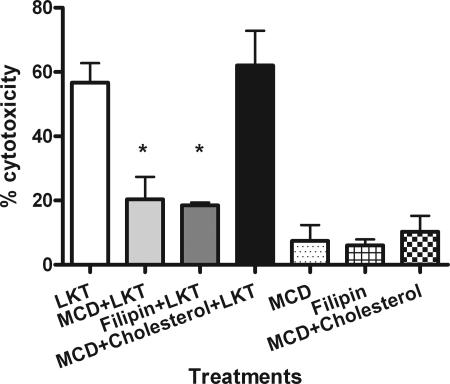
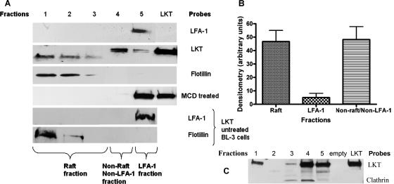
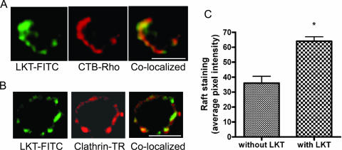
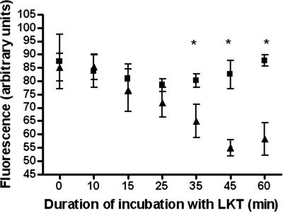
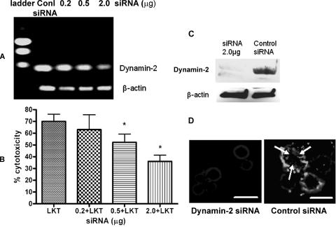
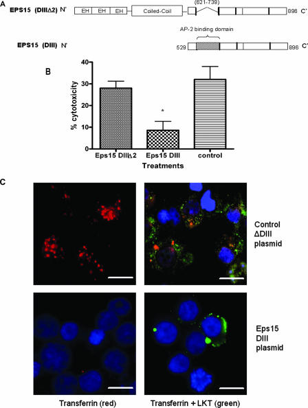
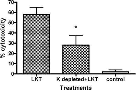
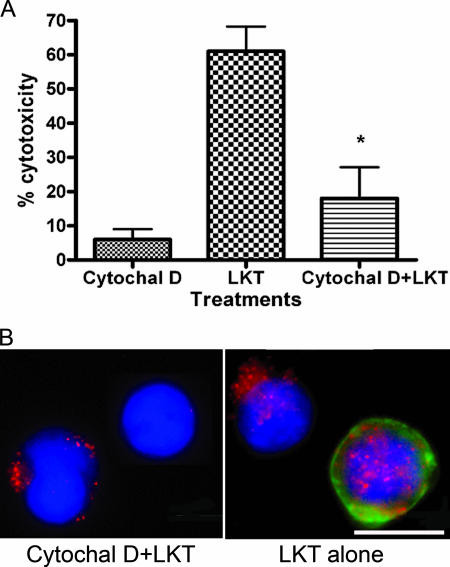
Mannheimia haemolytica is the principal bacterial pathogen of the bovine respiratory disease complex. Its most important virulence factor is a leukotoxin (LKT), which is a member of the RTX family of exotoxins produced by many gram-negative bacteria. Previous studies demonstrated that LKT binds to the beta(2)-integrin LFA-1 (CD11a/CD18) on bovine leukocytes, resulting in cell death. In this study, we demonstrated that depletion of lipid rafts significantly decreases LKT-induced bovine lymphoblastoid cell (BL-3) death. After binding to BL-3 cells, some of the LKT relocated to lipid rafts in an LFA-1-independent manner. We hypothesized that after binding to LFA-1 on BL-3 cells, LKT moves to lipid rafts and clathrin-coated pits via a dynamic process that results in LKT internalization and cytotoxicity. Knocking down dynamin-2 by small interfering RNA reduced both LKT internalization and cytotoxicity. Similarly, expression of dominant negative Eps15 protein expression, which is required for clathrin coat formation, reduced LKT internalization and LKT-mediated cytotoxicity to BL-3 cells. Finally, we demonstrated that inhibiting actin polymerization reduced both LKT internalization and LKT-mediated cytotoxicity. These results suggest that both lipid rafts and clathrin-mediated mechanisms are important for LKT internalization and cytotoxicity in BL-3 cells and illustrate the complex nature of LKT internalization by the cytoskeletal network.


Figures

FIG. 1.
FIG. 1.
Depletion and sequestration of lipid rafts in BL-3 cells inhibit LKT-mediated cytotoxicity. BL-3 cells were pretreated for 30 min at 37°C with either 5 mM MCD (to deplete cholesterol) or 4.5 μg/ml filipin (to sequester lipid rafts). Cells were then incubated with LKT (0.5 U) for 1 h. Cell viability was measured in a cytotoxicity assay as described in Materials and Methods. Some MCD-treated BL-3 cells were incubated with MCD-10 mM cholesterol (to reconstitute lipid rafts) before exposure to LKT. BL-3 cells incubated with MCD, filipin, or MCD-cholesterol alone were included as controls. Data are expressed as means and standard errors of the means of three separate experiments. An asterisk indicates that the P value is <0.05 for a comparison with BL-3 cells exposed to LKT alone.
FIG. 2.
FIG. 2.
LKT associates with detergent-resistant membrane fractions (lipid rafts) in BL-3 cells. A total of 106 BL-3 cells were incubated with LKT (0.5 U) for 30 min at 37°C and then lysed with TNE buffer. The lysates were centrifuged at 10,000 × g for 15 min. Detergent-resistant membrane fractions were isolated on Optiprep discontinuous gradients, and five fractions were collected from the top to the bottom (designated fractions 1 to 5). In panel A, protein was extracted from each fraction, and Western immunoblotting was performed for LKT, LFA-1, and flotillin. BL-3 cells depleted of cholesterol by MCD treatment and lipid raft fractions from non-LKT-treated BL-3 cells were included as controls and probed with anti-LFA-1 and anti-flotillin antibodies. In panel C, protein was extracted from each fraction, and Western immunoblotting was performed for LKT and clathrin light chain. This allowed us to characterize LFA-1 and LKT in raft and nonraft fractions. In panel B, the intensities of LKT bands in raft and nonraft fractions in panel A were quantified using LabWorks analysis software and are expressed as means and standard errors of the means of three separate experiments.
FIG. 3.
FIG. 3.
LKT colocalizes with lipid rafts and clathrin pits on BL-3 cells. A total of 106 BL-3 cells were incubated with LKT (0.5 U) for 30 min at 37°C. Cells were then fixed with 2% paraformaldehyde for 10 min, blocked with 3% bovine serum albumin for 20 min, and incubated for 1 h at 25°C with FITC-conjugated anti-LKT MAb, CTB-Rho (which binds to GM1 associated with lipid rafts), or Texas Red (TR)-conjugated anti-clathrin heavy chain antibody. Cells were visualized with a confocal microscope. Panel A shows a single cell in which LKT (green) colocalized with lipid rafts (red). Panel B shows colocalization of LKT with clathrin (red). In panel C, the mean lipid raft fluorescence intensities of images from 50 randomly selected cells were quantified for LKT-induced raft aggregation, using Image J software (NIH; http://rsb.info.nih.gov/ij). Bars = 10 μm.
FIG. 4.
FIG. 4.
Surface fluorescence for LKT on BL-3 cells decreased with time, while total fluorescence remained unchanged. A total of 106 BL-3 cells were incubated with LKT (0.5 U) for the indicated times (5 to 60 min) at 37°C and then fixed with 2% paraformaldehyde. Fixed BL-3 cells were stained with FITC-conjugated anti-LKT antibody for 60 min at 25°C, and the surface fluorescence was analyzed by flow cytometry (▴). To quantify total fluorescence of cells (▪), LKT-treated BL-3 cells were permeabilized with cold acetone (−20°C) for 10 min before labeling with FITC-conjugated anti-LKT antibody. The cells were then analyzed by flow cytometry. Fluorescence is expressed as the means ± standard errors of the means of three separate experiments. An asterisk indicates that the P value is <0.05.
FIG. 5.
FIG. 5.
siRNA knockdown of dynamin-2 reduces LKT-mediated cytotoxicity and internalization in BL-3 cells. Dynamin-2 was knocked down by transfecting BL-3 cells with siRNA (0.2, 0.5, or 2.0 μg) for 6 h, followed by further incubation in RPMI medium with 10% fetal bovine serum for 72 h. Control cells were transfected with a scrambled siRNA sequence for the same time period. To assess the degree of dynamin-2 RNA knockdown, RNA was extracted, and a one-step reverse transcription-PCR assay with dynamin-2-specific primers was performed. In panel A the products were visualized by electrophoresis in a 1% agarose gel. In panel B, dynamin-2 siRNA- or scrambled siRNA-transfected BL-3 cells (106) were incubated with LKT (0.5 U) for 1 h, and cytotoxicity was measured as described previously. The data are the means and standard errors of the means of five separate siRNA transfection experiments. An asterisk indicates that the P value is <0.05 for a comparison with the scrambled siRNA control. Panel C shows an immunoblot analysis of dynamin-2 knockdown from BL-3 cells transfected with 2 μg of dynamin-2 siRNA. In panel D, BL-3 cells transfected with dynamin-2 siRNA (2.0 μg) or with scrambled siRNA were fixed, permeabilized with cold acetone, blocked with 3% bovine serum albumin, and incubated with anti-LKT MAb. A goat anti-mouse IgG-Texas Red-conjugated secondary antibody was added, and the LKT signal was visualized by fluorescent microscopy at excitation and emission wavelengths of 595 and 614 nm, respectively. The arrows indicate internalized LKT. Bars = 10 μm. Conl, control.
FIG. 6.
FIG. 6.
Expression of a dominant negative Eps15 protein in BL-3 cells inhibits clathrin-dependent internalization of LKT and LKT-mediated cytotoxicity. Panel A is a schematic representation of the dominant negative mutant Eps15 protein (DIII) and the control Eps15 protein (DIIIΔ2) expressed in BL-3 cells. In the Eps15 (DIII) construct, the Eps15 homology domains (EH) and N-terminal coiled coil domains that are required for clathrin coat assembly have been deleted. In panel B, BL-3 cells were transfected with 6 μg of Eps15 DIII or the control Eps15 DIII Δ2 plasmid for 6 h at 37°C and then incubated with LKT (0.2 U) for 1 h at 37°C. Cytotoxicity was measured as described previously. The data are the means and standard errors of the means of three separate experiments. The asterisk indicates that the P value is <0.05. In panel C, BL-3 cells transfected with the Eps15 DIII or Eps15 DIII Δ2 plasmid were stained for cytoplasmic LKT and for transferrin (which is internalized by the clathrin-mediated pathway). BL-3 cells incubated with LKT were fixed, permeabilized, blocked with 3% bovine serum albumin, and incubated with FITC-conjugated anti-LKT MAb and rhodamine-conjugated transferrin. Images were visualized by confocal microscopy. The control plasmid-transfected BL-3 cells demonstrated internalization of transferrin (red) and LKT (green), while the Eps15 DIII plasmid-transfected BL-3 cells showed reduced internalization of LKT and virtually no signal from transferrin. Cells were counterstained with 4′,6′-diamidino-2-phenylindole (DAPI) nuclear stain (blue). The images in panel C show representative cells from three separate experiments that were performed. Bars = 10 μm.
FIG. 7.
FIG. 7.
Potassium depletion in BL-3 cells inhibits LKT-mediated cytotoxicity. BL-3 cells were depleted of K + by incubation in K+-free buffer for 60 min. Cells were then incubated with LKT, and cytotoxicity was measured. BL-3 cells treated with heat-inactivated LKT and K+-depleted BL-3 cells alone served as controls. The data are the means and standard errors of the means of three separate experiments performed. The asterisk indicates that the P value is <0.05 for a comparison with K+-depleted BL-3 cells. K+-depleted BL-3 cells demonstrated a significant reduction in tranferrin and LKT internalization (data not shown).
FIG. 8.
FIG. 8.
Cytochalasin D treatment inhibits LKT-mediated cytotoxicity and LKT internalization in BL-3 cells. In panel A, β-actin was depleted by treating BL-3 cells with cytochalasin D (Cytochal D) (2 μg/ml) for 60 min. Cells were then incubated with LKT, and cytotoxicity was assessed as described in the text. The data are as the means and standard errors of the means of three separate experiments performed. The asterisk indicates that the P value is <0.05 for a comparison with LKT alone. In panel B, intracellular staining for LKT was performed using an anti-LKT MAb and Texas Red-conjugated anti-mouse IgG. FITC-conjugated phalloidin was added to visualize actin filaments, and BL-3 cells were visualized by fluorescent microscopy. Cytochalasin D-treated cells showed a reduction in LKT internalization (red) and staining for β-actin (green). Cells were counterstained with DAPI nuclear stain (blue). BL-3 cells alone or treated with cytochalasin D were used as controls. Bar = 10 μm.

References

    1. Abrami, L., S. Liu, P. Cosson, S. H. Leppla, and F. G. van der Goot. 2003. Anthrax toxin triggers endocytosis of its receptor via a lipid raft-mediated clathrin-dependent process. J. Cell Biol. 160:321-328. - PMC - PubMed
    1. Ambagala, T. C., A. P. Ambagala, and S. Srikumaran. 1999. The leukotoxin of Pasteurella haemolytica binds to beta(2) integrins on bovine leukocytes. FEMS Microbiol. Lett. 179:161-167. - PubMed
    1. Atapattu, D. N., and C. J. Czuprynski. 2005. Mannheimia haemolytica leukotoxin induces apoptosis of bovine lymphoblastoid cells (BL-3) via a caspase-9-dependent mitochondrial pathway. Infect. Immun. 73:5504-5513. - PMC - PubMed
    1. Benmerah, A., M. Bayrou, N. Cerf-Bensussan, and A. Dautry-Varsat. 1999. Inhibition of clathrin-coated pit assembly by an Eps15 mutant. J. Cell Sci. 112:1303-1311. - PubMed
    1. Benmerah, A., C. Lamaze, B. Begue, S. L. Schmid, A. Dautry-Varsat, and N. Cerf-Bensussan. 1998. AP-2/Eps15 interaction is required for receptor-mediated endocytosis. J. Cell Biol. 140:1055-1062. - PMC - PubMed

Publication types

LinkOut - more resources