. 2007 Oct;75(10):4743-53.
doi: 10.1128/IAI.00687-07. Epub 2007 Aug 6.

Dynamics of Neisseria gonorrhoeae attachment: microcolony development, cortical plaque formation, and cytoprotection

Affiliations

Dynamics of Neisseria gonorrhoeae attachment: microcolony development, cortical plaque formation, and cytoprotection

Dustin L Higashi et al. Infect Immun. 2007 Oct.

Abstract

Neisseria gonorrhoeae is the bacterium that causes gonorrhea, a major sexually transmitted disease and a significant cofactor for human immunodeficiency virus transmission. The retactile N. gonorrhoeae type IV pilus (Tfp) mediates twitching motility and attachment. Using live-cell microscopy, we reveal for the first time the dynamics of twitching motility by N. gonorrhoeae in its natural environment, human epithelial cells. Bacteria aggregate into microcolonies on the cell surface and induce a massive remodeling of the microvillus architecture. Surprisingly, the microcolonies are motile, and they fuse to form progressively larger structures that undergo rapid reorganization, suggesting that bacteria communicate with each other during infection. As reported, actin plaques form beneath microcolonies. Here, we show that cortical plaques comigrate with motile microcolonies. These activities are dependent on pilT, the Tfp retraction locus. Cultures infected with a pilT mutant have significantly higher numbers of apoptotic cells than cultures infected with the wild-type strain. Inducing pilT expression with isopropyl-beta-D-thiogalactopyranoside partially rescues cells from infection-induced apoptosis, demonstrating that Tfp retraction is intrinsically cytoprotective for the host. Tfp-mediated attachment is therefore a continuum of microcolony motility and force stimulation of host cell signaling, leading to a cytoprotective effect.


Figures

FIG. 1.
FIG. 1.
Microcolony fusion and realignment. Time-lapse images of three fusing microcolonies (see Video S1 in the supplemental material) of A431 cells infected with wt MS11. The time stamps on the lower right corners indicate h:min postinfection. (A and B) Three microcolonies (arrowheads) before fusion. (C, D, E, and F) The same microcolonies undergoing fusion and realignment. Scale bar, 10 μm.
FIG. 2.
FIG. 2.
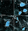
Shapes of wt and pilT mutant microcolonies and morphologies of host cell microvilli. Shown are SEM images of A431 cells incubated for 3 h with medium (A), wt MS11 (B and C), or MS11pilT (D). Bacteria are color enhanced (blue) to provide added contrast with the epithelial cells (gray). (B and C) Domed and bilobed shapes of wt microcolonies. The arrowheads point to stretched and elongated microvilli beneath microcolonies. Other microvilli appear to wrap around individual diplococci at the peripheries of, or away from, microcolonies. (A) Morphology of the mock-infected cell surface. (D) Irregular shape of MS11pilT clusters and lack of microvillus remodeling. Scale bar, 10 μm.
FIG. 3.
FIG. 3.
Movement of wt and pilT microcolonies during infection. (A) Trajectories of the movements of wt MS11 (left) and MS11pilT (right) microcolonies on A431 epithelial cells over a 5.5-hour infection (see Materials and Methods for details). The color code in each track shows the time of image acquisition. Blue represents the beginning of image acquisition. White represents the end of image acquisition. Color bar, 5.5 h. (B) Distances traveled by microcolonies from the point of origin expressed as a function of time (each unit represents 2 min) for wt MS11 (left) and MS11pilT (right).
FIG. 4.
FIG. 4.
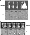
Positions of actin-GFP plaques relative to a moving microcolony. Time-lapse images (see Video S2 in the supplemental material) were compiled to show two different actin-GFP-expressing A431 epithelial cells with infecting wt MS11 microcolonies. The time stamps indicate the times of image acquisition in hr:min:s. (A) Clustering of actin-GFP at the site of entry of a microcolony. (Top) Successive FITC images showing actin-GFP clustering in the cell. Directly below each FITC image is the corresponding DIC image, presented to show the position of the microcolony. Frame i was acquired prior to entry of the microcolony onto the cell. The arrow points to the entry site of the microcolony; its position in all frames is invariant. (B) Movement of actin-GFP plaque relative to the position of a motile microcolony. (Top) Successive FITC images of another infected cell showing the positions of the actin-GFP plaque. Directly below each FITC image is the corresponding DIC image showing the positions of the microcolony. The arrow in each panel points to the original position of the microcolony; its position in all frames is invariant. Scale bar, 10 μm.
FIG. 5.
FIG. 5.
Morphology of actin-GFP-expressing A431 cells infected with MS11pilT (A) or treated with the apoptosis-inducing agent STS (B). The images in panels A and B are taken from successive video images (see Videos S3 and S4, respectively, in the supplemental material). The top rows in panels A and B show FITC images of A431 cells. The DIC image corresponding to each FITC image appears immediately below. Both sets of images were acquired over a 12.5-min period. The arrows indicate apoptotic bodies. Scale bar, 11 μm.
FIG. 6.
FIG. 6.
Immunodetection of PAR in infected A431 cells. A431 cells were mock infected with medium (A), infected with wt MS11 (B) or MS11pilT (C) for 5.5 h, or treated with STS for 4.5 h (D). Samples were fixed and stained with antibodies to PAR (green) and DAPI (blue) to visualize nuclei. All images were acquired on a Nikon Microphot-FX at the same magnification. PAR and DAPI signals are superimposed in all images.
FIG. 7.
FIG. 7.
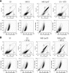
Dual-fluorescence flow cytometry analysis of cell death in N. gonorrhoeae-infected cultures. (A) A431 cells were mock infected with medium (UI), infected with wt MS11 or MS11pilT for 5.5 h, or treated with STS for 4.5 h and analyzed by flow cytometry for YO-PRO-1 and PI staining (top row). (B) A431 cells were infected with MS11pilTi in the presence or absence of IPTG. Parallel mock-infected (UI) cultures were treated with IPTG or left untreated, as indicated (see Materials and Methods), and analyzed by flow cytometry for YO-PRO-1 and PI staining. The top rows in panels A and B contain bivariate dot plots of cells in each sample stained with these dyes. The number in gate A indicates the percentage of YO-PRO-1+ PI cells in the population. The number in gate D indicates the percentage of YO-PRO-1+ PI+ cells. Below each sample is its corresponding scattergram (lower rows). The gated populations within each scattergram were used to plot PI fluorescence in relation to YO-PRO-1.

Similar articles

Cited by

References

    1. Achtman, M., M. Neibert, B. A. Crowe, W. Strittmatter, B. Kusecek, E. Weyse, M. J. Walsh, B. Slawig, G. Morelli, A. Moll, et al. 1988. Purification and characterization of eight class 5 outer membrane protein variants from a clone of Neisseria meningitidis serogroup A. J. Exp. Med. 168:507-525. - PMC - PubMed
    1. Aukema, K. G., E. M. Kron, T. J. Herdendorf, and K. T. Forest. 2005. Functional dissection of a conserved motif within the pilus retraction protein PilT. J. Bacteriol. 187:611-618. - PMC - PubMed
    1. Ayala, P., J. S. Wilbur, L. M. Wetzler, J. A. Tainer, A. Snyder, and M. So. 2005. The pilus and porin of Neisseria gonorrhoeae cooperatively induce Ca2+ transients in infected epithelial cells. Cell. Microbiol. 7:1736-1748. - PubMed
    1. Belmokhtar, C. A., J. Hillion, and E. Segal-Bendirdjian. 2001. Staurosporine induces apoptosis through both caspase-dependent and caspase-independent mechanisms. Oncogene 20:3354-3362. - PubMed
    1. Bernard, B., T. Fest, J. L. Pretet, and C. Mougin. 2001. Staurosporine-induced apoptosis of HPV positive and negative human cervical cancer cells from different points in the cell cycle. Cell Death. Differ. 8:234-244. - PubMed

Publication types