. 2007 Aug 13;178(4):575-81.
doi: 10.1083/jcb.200612022. Epub 2007 Aug 6.

p32 is a novel mammalian Lgl binding protein that enhances the activity of protein kinase Czeta and regulates cell polarity

Affiliations

p32 is a novel mammalian Lgl binding protein that enhances the activity of protein kinase Czeta and regulates cell polarity

Carl U Bialucha et al. J Cell Biol. .

Abstract

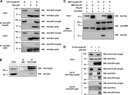
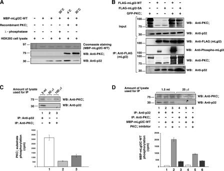
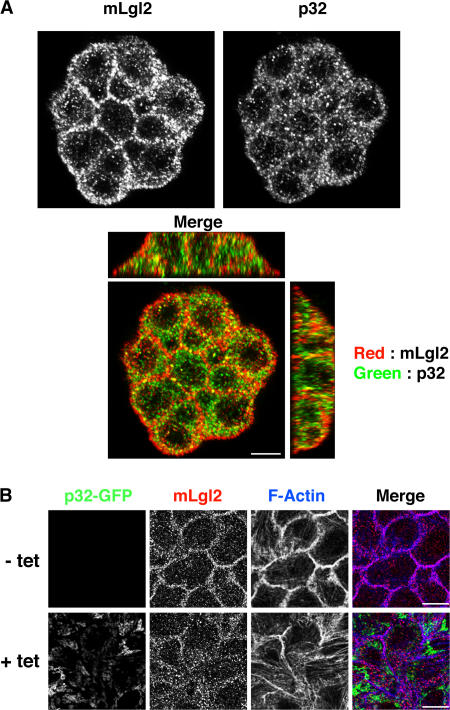
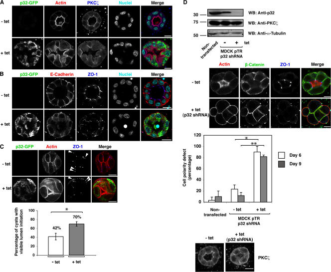
Lgl (lethal giant larvae) plays an important role in cell polarity. Atypical protein kinase C (aPKC) binds to and phosphorylates Lgl, and the phosphorylation negatively regulates Lgl activity. In this study, we identify p32 as a novel Lgl binding protein that directly binds to a domain on mammalian Lgl2 (mLgl2), which contains the aPKC phosphorylation site. p32 also binds to PKCzeta, and the three proteins form a transient ternary complex. When p32 is bound, PKCzeta is stimulated to phosphorylate mLgl2 more efficiently. p32 overexpression in Madin-Darby canine kidney cells cultured in a 3D matrix induces an expansion of the actin-enriched apical membrane domain and disrupts cell polarity. Addition of PKCzeta inhibitor blocks apical actin accumulation, which is rescued by p32 overexpression. p32 knockdown by short hairpin RNA also induces cell polarity defects. Collectively, our data indicate that p32 is a novel regulator of cell polarity that forms a complex with mLgl2 and aPKC and enhances aPKC activity.


Figures

Figure 1.
Figure 1.
p32 is a novel mLgl2 binding protein. (A) Reciprocal coimmunoprecipitation of mLgl2 and p32. GFP-mLgl2-WT and Myc-p32 were expressed in HEK293 cells, and immunoprecipitation was performed by using either anti-GFP or anti-Myc antibody, followed by Western blotting with anti-GFP and anti-Myc antibodies. (B) Coimmunoprecipitation of endogenous mLgl2 and p32 proteins. Immunoprecipitation was performed with anti-mLgl2 antibody from HEK293 cell lysate, followed by Western blotting using anti-mLgl2 and anti-p32 antibodies. (C) Direct interaction between p32 and mLgl2. Recombinant MBP-mLgl2C-WT and His-p32 proteins were incubated and pulled down using amylose resin beads, followed by Western blotting using anti-MBP and anti-His antibodies. As controls, MBP-Rac-WT and His-Hakai were used. (D) Double immunoprecipitation of mLgl2 and p32 to analyze complex formation with PKCζ. FLAG-mLgl2-WT and p32-GFP were expressed in HEK293 cells. An initial immunoprecipitation was performed using anti-FLAG antibody, followed by elution of immunoprecipitates with FLAG peptide. The eluates were subjected to a second immunoprecipitation using anti-GFP antibody. The first and second immunoprecipitates were examined by Western blotting using anti-FLAG, anti-GFP, and anti-PKCζ antibodies.
Figure 2.
Figure 2.
p32 interacts with mLgl2 transiently and enhances PKCζ activity. (A and B) The interaction of p32 with mLgl2 was examined in the presence or absence of PKCζ under different mLgl2 phosphorylation conditions. (A) MBP-mLgl2C-WT was incubated with PKCζ or λ-phosphatase at 30 or 4°C, followed by incubation with HEK293 cell lysates and pull-down assay using amylose resin beads. PKCζ and endogenous p32 bound to MBP-mLgl2C-WT were examined by Western blotting using anti-PKCζ and anti-p32 antibodies, respectively. The membrane was stained with Coomassie brilliant blue to visualize MBP-mLgl2C-WT. (B) FLAG-mLgl2-WT or -SA was expressed in HEK293 cells with or without coexpression of GFP-PKCζ. Immunoprecipitation was performed using anti-FLAG antibody, followed by Western blotting with anti-FLAG, anti–phospho-mLgl2, anti-PKCζ, and anti-p32 antibodies. (C) Binding of p32 enhances the activity of PKCζ to phosphorylate a PKCζ peptide substrate. PKCζ was immunoprecipitated from the indicated amounts of HEK293 cell lysate by using either anti-p32 or anti-PKCζ antibody. In vitro kinase assays were performed by incubating immunoprecipitates with PKCζ peptide substrate and γ-[32P]ATP. Peptide substrates were then collected by incubation with streptavidin beads, and the radioactivity of the beads was measured. Without peptide substrate, negligible radioactivity was measured using streptavidin beads alone. (D) Binding of p32 enhances the activity of PKCζ to phosphorylate mLgl2. Immunoprecipitations and in vitro kinase assays were performed as described, except that MBP-mLgl2C-WT was used as PKCζ substrate, and incubations were performed in the presence or absence of a PKCζ inhibitor. (C and D) The radioactive count by control IgG immunoprecipitate was subtracted from that by p32 and PKCζ immunoprecipitates. Phosphorylation reactions were duplicated for each experiment, and the results shown are representative of three independent experiments. Error bars indicate mean ± SD.
Figure 3.
Figure 3.
p32 partially colocalizes with mLgl2, and overexpression of p32 affects the localization of mLgl2 in MDCK cells. (A) Immunofluorescence analysis of endogenous mLgl2 and p32 proteins in MDCK cells. (B) Immunofluorescence analysis of p32-GFP, mLgl2, and F-actin in MDCK cells stably expressing p32-GFP in a tetracycline-inducible manner. These results are representative of results from two independent clones of p32-GFP MDCK cells. Bars, 10 μm.
Figure 4.
Figure 4.
p32 expression level affects cell polarity of MDCK cells in 3D culture. (A and B) Immunofluorescence analysis of cell polarity markers in p32-GFP MDCK cell cysts. MDCK cells stably expressing p32-GFP in a tetracycline-inducible manner were seeded in a collagen gel. Cells were either treated or not treated with tetracycline to induce p32-GFP expression. (A) Immunostaining was performed using anti-GFP and anti-PKCζ antibodies. Actin was visualized with TRITC-labeled phalloidin. Nuclei were stained with Hoechst dye. (B) Immunostaining was performed using anti-GFP, anti-E-cadherin, and anti–ZO-1 antibodies. (C) Cell polarity defects in p32-overexpressing MDCK cells are linked to PKCζ activation. A cell-permeable PKCζ pseudosubstrate inhibitor was added to noninduced or p32-GFP–overexpressing cells. Immunostaining was performed using anti-GFP and –ZO-1 antibodies. Arrowheads indicate ectopic localizations of ZO-1. These results are representative of results from two independent clones of p32-GFP MDCK cells. *, P < 0.05. (D) A knockdown of p32 induces cell polarity defects in p32 shRNA–expressing MDCK cells. (top) Western blotting with anti-p32, anti-PKCζ, or anti–α-tubulin antibody was performed using cell lysates from parental or p32 shRNA cells. (second and bottom panels) Immunofluorescence analysis of cell polarity markers in p32 shRNA MDCK cell cysts. Immunostaining was performed using TRITC-labeled phalloidin and anti–β-catenin, anti–ZO-1, and anti-PKCζ antibodies. These results are representative of results from two independent clones of p32 shRNA MDCK cells. (third panel) For statistical analysis, 20 cysts were examined in each experiment at day 6 or 9 after seeding, and the results represent the means ± SD of three independent experiments. *, P < 0.0015; **, P < 0.0005. Cysts were analyzed by confocal microscopy. Bars, 10 μm.

References

    1. Betschinger, J., K. Mechtler, and J.A. Knoblich. 2003. The Par complex directs asymmetric cell division by phosphorylating the cytoskeletal protein Lgl. Nature. 422:326–330. - PubMed
    1. Betschinger, J., F. Eisenhaber, and J.A. Knoblich. 2005. Phosphorylation- induced autoinhibition regulates the cytoskeletal protein Lethal (2) giant larvae. Curr. Biol. 15:276–282. - PubMed
    1. Bilder, D. 2004. Epithelial polarity and proliferation control: links from the Drosophila neoplastic tumor suppressors. Genes Dev. 18:1909–1925. - PubMed
    1. Bilder, D., M. Li, and N. Perrimon. 2000. Cooperative regulation of cell polarity and growth by Drosophila tumor suppressors. Science. 289:113–116. - PubMed
    1. Chalmers, A.D., M. Pambos, J. Mason, S. Lang, C. Wylie, and N. Papalopulu. 2005. aPKC, Crumbs3 and Lgl2 control apicobasal polarity in early vertebrate development. Development. 132:977–986. - PubMed

Publication types