. 2007 Oct;27(19):6806-17.
doi: 10.1128/MCB.01036-07. Epub 2007 Aug 6.

Elucidation of a C-rich signature motif in target mRNAs of RNA-binding protein TIAR

Affiliations

Elucidation of a C-rich signature motif in target mRNAs of RNA-binding protein TIAR

Henry S Kim et al. Mol Cell Biol. 2007 Oct.

Abstract

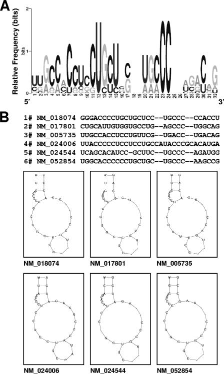
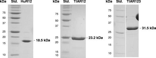
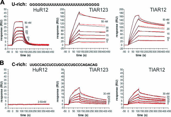
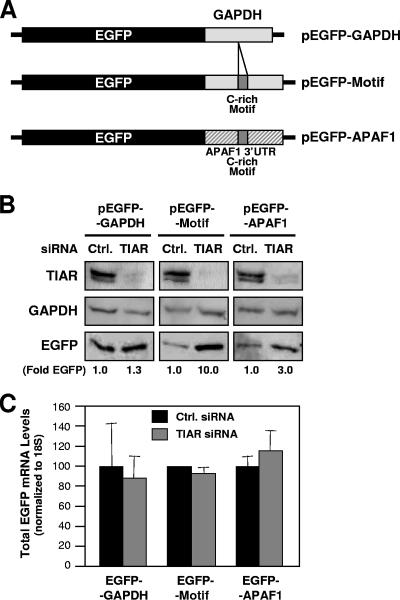
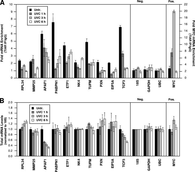
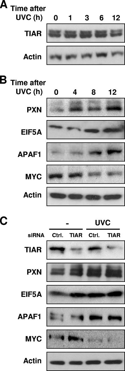
The RNA-binding protein TIAR (related to TIA-1 [T-cell-restricted intracellular antigen 1]) was shown to associate with subsets of mRNAs bearing U-rich sequences in their 3' untranslated regions. TIAR can function as a translational repressor, particularly in response to cytotoxic agents. Using unstressed colon cancer cells, collections of mRNAs associated with TIAR were isolated by immunoprecipitation (IP) of (TIAR-RNA) ribonucleoprotein (RNP) complexes, identified by microarray analysis, and used to elucidate a common signature motif present among TIAR target transcripts. The predicted TIAR motif was an unexpectedly cytosine-rich, 28- to 32-nucleotide-long element forming a stem and a loop of variable size with an additional side loop. The ability of TIAR to bind an RNA oligonucleotide with a representative C-rich TIAR motif sequence was verified in vitro using surface plasmon resonance. By this analysis, TIAR containing two or three RNA recognition domains (TIAR12 and TIAR123) showed low but significant binding to the C-rich sequence. In vivo, insertion of the C-rich motif into a heterologous reporter strongly suppressed its translation in cultured cells. Using this signature motif, an additional approximately 2,209 UniGene targets were identified (2.0% of the total UniGene database). A subset of specific mRNAs were validated by RNP IP analysis. Interestingly, in response to treatment with short-wavelength UV light (UVC), a stress agent causing DNA damage, each of these target mRNAs bearing C-rich motifs dissociated from TIAR. In turn, expression of the encoded proteins was elevated in a TIAR-dependent manner. In sum, we report the identification of a C-rich signature motif present in TIAR target mRNAs whose association with TIAR decreases following exposure to a stress-causing agent.


Figures

FIG. 1.
FIG. 1.
Sequence and structure of the predicted TIAR motif, as identified among TIAR-bound transcripts. (A) Probability matrix (graphic logo) of the TIAR motif indicating the relative frequency of finding each residue at each position within the motif, as elucidated from the array-derived experimental data set. (B) Secondary structure of six representative examples of the TIAR motif in specific mRNAs; the corresponding RefSeq accession numbers names are shown.
FIG. 2.
FIG. 2.
Recombinant proteins used in in vitro binding assays. The construction of plasmids to express recombinant proteins comprising RRM1 and RRM2 or all three RRMs (HuR12, TIAR123, and TIAR12) was previously described (10). Proteins were expressed in bacteria and purified to homogeneity (details in Materials and Methods). Std., protein standard, with molecular mass (kDa) indicated.
FIG. 3.
FIG. 3.
Kinetic analysis of the interactions of TIAR12, TIAR123, and HuR12 proteins with U-rich and C-rich RNAs. The binding of TIAR123, TIAR12, and HuR12 to a U-rich or a C-rich RNA (28-mer each) is shown. Biotinylated RNA was captured on SA-coated sensor chips, and increasing concentrations of protein were injected over the surface. Injections were performed for 120 s (association phase), followed by a 300-s flow of running buffer to assess dissociation. The experiments were conducted in duplicate and showed good overlap. The red lines represent the binding responses for injections of protein analyte at specified concentrations (nM) over the RNA surface. The kinetic data were fit by a 1:1 Langmuir binding model which describes monovalent analyte binding to a single site on the immobilized ligand. Mass transport effects were not evident. The black curves superimposed on top of the sensorgrams represent the model fitted curves. The association and dissociation rate constants (Ka and Kd, respectively) were determined simultaneously as global fitting parameters from which KD was determined. The resulting parameter values are given in Table 2.
FIG. 4.
FIG. 4.
Functional assessment of the C-rich motif using a heterologous reporter. (A) Plasmid pEGFP-GAPDH contains a 200-bp fragment of the GAPDH 3′UTR after the EGFP coding region; plasmid pEGFP-APAF1 contains nucleotide positions 5401 to 5825 of APAF1 (NM_013229), including one hit of the APAF1 3′UTR C-rich motif (positions 5599 to 5626); plasmid pEGFP-Motif contains a 28-bp insert (the consensus C-rich motif) within the GAPDH 3′UTR (Materials and Methods). (B) Western blot analysis of TIAR, loading control GAPDH, and reporter protein EGFP expression levels in RKO cells that had been transfected 48 h earlier with either control (Ctrl.) or TIAR-directed siRNAs. Signals (representative of five independent experiments) were quantified by densitometry and shown as EGFP levels (fold) in TIAR siRNA compared with control siRNA. (C) The levels of chimeric EGFP-GAPDH, EGFP-APAF1, and EGFP-Motif mRNAs were measured in each transfection group (B) by RT-qPCR analysis. Data are shown as the means and standard errors of the mean of three independent experiments.
FIG. 5.
FIG. 5.
Analysis of the binding and whole-cell levels of predicted TIAR target mRNAs in untreated and UVC-irradiated cells. (A) The association of endogenous TIAR with endogenous putative target mRNAs was tested using lysates prepared from RKO cells that were either left without treatment (Untr. [0 h]) or were irradiated with 25 J/m2 UVC and collected 1, 3, or 6 h afterwards. Anti-TIAR or IgG antibodies were used in IP reactions followed by the analysis of predicted target transcripts by RT-qPCR analysis of the IP material. Neg., negative control transcripts 18S rRNA and housekeeping GAPDH and UBC mRNAs; Pos., positive control MYC mRNA, a known TIAR target (32). Numbers on the right y axis indicate MYC mRNA enrichment (TIAR/IgG). (B) Total RNA was extracted from cells that were processed as described for panel A. The whole-cell levels of each mRNA were calculated and normalized to 18S rRNA levels. Data show means and standard deviations (A and B).
FIG. 6.
FIG. 6.
Expression levels of proteins encoded by predicted TIAR target mRNAs in untreated and UVC-irradiated RKO cells. (A) Western blot analysis of TIAR expression levels in whole-cell lysates (15 μg per lane) that were prepared at the times indicated after UVC irradiation. (B) Western blot analysis of the expression levels of PXN, EIF5A, and APAF1 (encoded by TIAR dissociation target mRNAs), positive control MYC, and loading control β-actin. Whole-cell lysates (10 μg per lane) were prepared at the times shown following UVC irradiation. Shown are representative Western blots from three independent experiments. (C) Western blot analysis of the expression of TIAR, PXN, EIF5A, APAF1, MYC, and β-actin in cells that were transfected with either control (Ctrl.) or TIAR siRNAs. Forty-eight hours later, cells were left untreated or treated with UVC and collected after an additional 8 h. Data are representative of three independent experiments.

References

    1. Anderson, P., and N. Kedersha. 2002. Stressful initiations. J. Cell Sci. 115:3227-3234. - PubMed
    1. Anderson, P., and N. Kedersha. 2002. Visibly stressed: the role of eIF2, TIA-1, and stress granules in protein translation. Cell Stress Chaperones 7:213-221. - PMC - PubMed
    1. Anderson, P., and N. Kedersha. 2007. On again, off again: the SRC-3 transcriptional coactivator moonlights as a translational corepressor. Mol. Cell 25:796-797. - PubMed
    1. Antic, D., and J. D. Keene. 1997. Embryonic lethal abnormal visual RNA-binding proteins involved in growth, differentiation, and posttranscriptional gene expression. Am. J. Hum. Genet. 61:273-278. - PMC - PubMed
    1. Antic, D., N. Lu, and J. D. Keene. 1999. ELAV tumor antigen, Hel-N1, increases translation of neurofilament M mRNA and induces formation of neurites in human teratocarcinoma cells. Genes Dev. 13:449-461. - PMC - PubMed

Publication types

MeSH terms

LinkOut - more resources