. 2007 Oct;27(19):6659-68.
doi: 10.1128/MCB.00205-07. Epub 2007 Aug 6.

Polysialic acid-directed migration and differentiation of neural precursors are essential for mouse brain development

Affiliations

Polysialic acid-directed migration and differentiation of neural precursors are essential for mouse brain development

Kiyohiko Angata et al. Mol Cell Biol. 2007 Oct.

Abstract

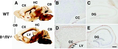
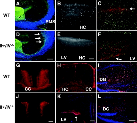
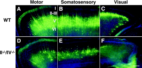
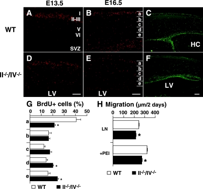
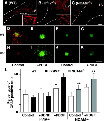
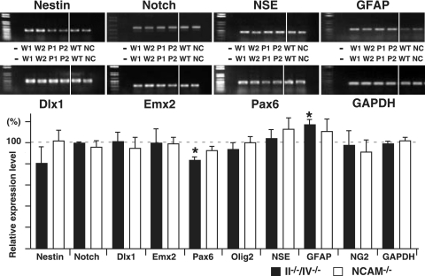
Polysialic acid, which is synthesized by two polysialyltransferases, ST8SiaII and ST8SiaIV, plays an essential role in brain development by modifying the neural cell adhesion molecule (NCAM). It is currently unclear how polysialic acid functions in different processes of neural development. Here we generated mice doubly mutant in both ST8SiaII and ST8SiaIV to determine the effects of loss of polysialic acid on brain development. In contrast to NCAM-deficient, ST8SiaII-deficient, or ST8SiaIV-deficient single mutant mice, ST8SiaII and ST8SiaIV double mutants displayed severe defects in anatomical organization of the forebrain associated with apoptotic cell death. Loss of polysialic acid affected both tangential and radial migration of neural precursors during cortical development, resulting in aberrant positioning of neuronal and glial cells. Glial cell differentiation was aberrantly increased in vivo and in vitro in the absence of polysialic acid. Consistent with these findings, polysialic acid-deficient mice exhibited increased expression of the glial cell marker glial fibrillary acidic protein and a decrease in expression of Pax6, a transcription factor regulating neural cell migration. These results indicate that polysialic acid regulates cell migration and differentiation of neural precursors crucial for brain development.


Figures

FIG. 1.
FIG. 1.
Apoptotic cell death in the cortex of polysialic acid-deficient mice. (A) Vibratome sections showing sagittal views of 1-month-old WT (upper) and II−/−/IV−/− (lower) mice. CB, cerebellum; CX, cerebral cortex; HC, hippocampus; LV, lateral ventricle; OB, olfactory bulb. (B to E) Apoptotic cells (brown) in sagittal sections of 1-month-old WT (B and C) and II−/−/IV−/− (D and E) mice were detected. Nuclei were stained by cresyl violet. The cerebral cortex (B and D) and hippocampus (C and E) are shown. Many cells undergo apoptosis in the SVZ of double mutant mice (arrows in panel D). CC, corpus callosum; DG, dentate gyrus. Bar, 0.2 mm.
FIG. 2.
FIG. 2.
Polysialic acid is required for cerebral cortex development. (A and D) Calretinin expression in olfactory neuron precursors (green) of P10 mice. Arrows indicate accumulated cells near the SVZ. (B and E) CaBP (white) marks GABAergic interneurons in a 1-month-old cortex. (C, F, G, and J) Nonphosphorylated neurofilament (red) marks pyramidal cells in cortical layers II and III and V and VI in sagittal sections (C and F) and coronal sections (G and J) of 1-month-old mice. In II−/−/IV−/− mice, neurofilament-expressing cells remained near the ventricular zone (arrow in panel F). Nissl substance staining (green) shows the distribution of neurons (C and F). (H, I, K, and L) Distribution of glial cells expressing GFAP (red) in coronal sections of the cortex (H and K) and hippocampal sagittal sections (I and L) of 1-month-old mice. Note that the number of astrocyte-like cells was decreased in the hippocampus of II−/−/IV−/− mice (L). Panels A to C and G to I show WT mice, and panels D to F and J to L show II−/−/IV−/− mice. CC, corpus callosum; DG, dentate gyrus; HC, hippocampus; LV, lateral ventricle. Bars, 0.2 mm.
FIG. 3.
FIG. 3.
Lack of polysialic acid affects lamination of pyramidal cells in the cerebral cortex. Images show YFP expression (green) in Thy1-YFP mice and Hoechst staining (blue) in 1-month-old WT (A to C) and II−/−/IV−/− (D to F) mice. Pyramidal neurons in layer V of the motor (A and D), somatosensory (B and E), and visual (C and F) cortex are shown. Bars, 0.2 mm. Note that in polysialic acid-deficient mice the number of pyramidal cells was decreased and the number of neuronal projections was decreased and not as extensive as in WT mice.
FIG. 4.
FIG. 4.
Effect of loss of polysialic acid on cell migration. (A, B, D, and E) BrdU-positive cells (red) at P0 are shown after BrdU injection at E13.5 (A and D) or E16.5 (B and E). Results obtained from littermates including both WT and mutant mice in each experiment were compared. Note that the number of BrdU-positive cells in the cerebral cortex was almost equivalent between wild-type and knockout mice. (C and F) P0 sections were stained with Ki67. (G) Distribution of BrdU-positive cells at P0 from the E16.5 injection was analyzed in four mice of each genotype. The cortex was equally divided into five bins (a to e in panels B and E), and BrdU-positive cells were counted in each bin. Forty percent of BrdU-labeled cells in the cortex reached bin a in WT mice, while 20% reached the same destination in II−/−/IV−/− mice, a statistically significant difference. Error bars indicate the standard deviations. *, P < 0.002. (H) Migration of neurosphere cells cultured for 2 days in 12-well plates coated with laminin (LN) alone or plus polyethylenimine (PEI). Neurosphere cells from II−/−/IV−/− mice migrated significantly more slowly than did wild-type cells. Error bars indicate the standard errors of the means. **, P < 0.001. Panels A, B, and C show WT mice, and panels D, E, and F show II−/−/IV−/− mice. HC, hippocampus; LV, lateral ventricle. Bars, 0.2 mm.
FIG. 5.
FIG. 5.
Roles of polysialic acid in cell differentiation. (A to C) Distribution of glial cells expressing GFAP (red) in the RMS of wild-type (A), II−/−/IV−/− (B), and NCAM−/− (C) mice. The RMS defined by Hoechst staining (not shown) is indicated by dotted lines. Enlarged images of GFAP-positive cells are shown in insets in panels A and B. LV, lateral ventricle. (D to K) Neurosphere cells regrown after dissociation were induced to differentiate by culturing without FGF2 (D, F, H, and J) but with PDGF-AA (E, G, I, and K) and stained for markers of neurons (β-III tubulin; red in panels D and H) and astrocytes (GFAP; green). Panels D to G show WT mice, panels H and I show II−/−/IV−/− mice, and panels J and K show NCAM−/− mice. (L) The proportion of astrocytes in the total number of differentiated cells is shown (four for each neurosphere clone for II−/−/IV−/− mice and two for each clone for NCAM−/− mice). Error bars indicate the standard deviations. *, P < 0.005; **, P < 0.01. Bar, 0.2 mm.
FIG. 6.
FIG. 6.
Comparative RT-PCR analysis of NCAM- and polysialic acid-deficient mice. (Upper) Total RNA from forebrain was prepared at P0 from WT and mutant mice from the same litter, and expression levels of various genes were analyzed using specific primers by RT-PCR. Photographs of amplified fragments are shown. −, without reverse transcriptase; W1, W2, and WT are WT mice, P1 and P2 are II−/−/IV−/− mice, and NC are NCAM−/− mice. (Lower) Amplified fragments were analyzed densitometrically and compared between WT and knockout mice in the same litter. At least two different mice of each genotype were used for statistical analysis. Error bars indicate standard deviations. *, P < 0.002; **, P < 0.005.

References

    1. Amoureux, M. C., B. A. Cunningham, G. M. Edelman, and K. L. Crossin. 2000. N-CAM binding inhibits the proliferation of hippocampal progenitor cells and promotes their differentiation to a neuronal phenotype. J. Neurosci. 15:3631-3640. - PMC - PubMed
    1. Angata, K., D. Chan, J. Thibault, and M. Fukuda. 2004. Molecular dissection of the ST8Sia IV polysialyltransferase. Distinct domains are required for neural cell adhesion molecule recognition and polysialylation. J. Biol. Chem. 279:25883-25890. - PubMed
    1. Angata, K., and M. Fukuda. 2003. Polysialyltransferases: major players in polysialic acid synthesis on the neural cell adhesion molecule. Biochimie 85:195-206. - PubMed
    1. Angata, K., J. M. Long, O. Bukalo, W. Lee, A. Dityatev, A. Wynshaw-Boris, M. Schachner, M. Fukuda, and J. D. Marth. 2004. Sialyltransferase ST8Sia-II assembles a subset of polysialic acid that directs hippocampal axonal targeting and promotes fear behavior. J. Biol. Chem. 279:32603-32613. - PubMed
    1. Berglund, E. O., K. K. Murai, B. Fredette, G. Sekerkova, B. Marturano, L. Weber, E. Mugnaini, and B. Ranscht. 1999. Ataxia and abnormal cerebellar microorganization in mice with ablated contactin gene expression. Neuron 24:739-750. - PubMed

Publication types

MeSH terms