. 2007 Sep 3;204(9):2023-30.
doi: 10.1084/jem.20070064. Epub 2007 Aug 6.

An antigen-specific pathway for CD8 T cells across the blood-brain barrier

Affiliations

An antigen-specific pathway for CD8 T cells across the blood-brain barrier

Ian Galea et al. J Exp Med. .

Abstract

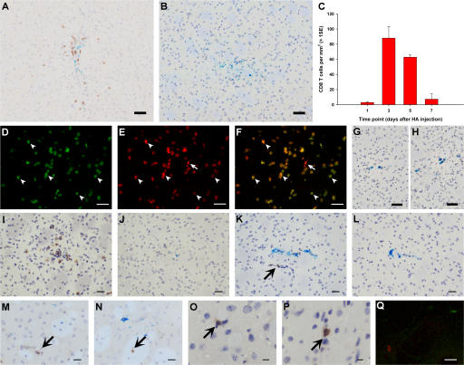
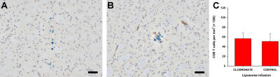
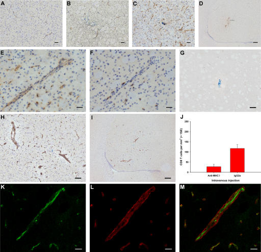
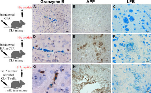
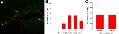
CD8 T cells are nature's foremost defense in encephalitis and brain tumors. Antigen-specific CD8 T cells need to enter the brain to exert their beneficial effects. On the other hand, traffic of CD8 T cells specific for neural antigen may trigger autoimmune diseases like multiple sclerosis. T cell traffic into the central nervous system is thought to occur when activated T cells cross the blood-brain barrier (BBB) regardless of their antigen specificity, but studies have focused on CD4 T cells. Here, we show that selective traffic of antigen-specific CD8 T cells into the brain occurs in vivo and is dependent on luminal expression of major histocompatibility complex (MHC) class I by cerebral endothelium. After intracerebral antigen injection, using a minimally invasive technique, transgenic CD8 T cells only infiltrated the brain when and where their cognate antigen was present. This was independent of antigen presentation by perivascular macrophages. Marked reduction of antigen-specific CD8 T cell infiltration was observed after intravenous injection of blocking anti-MHC class I antibody. These results expose a hitherto unappreciated route by which CD8 T cells home onto their cognate antigen behind the BBB: luminal MHC class I antigen presentation by cerebral endothelium to circulating CD8 T cells. This has implications for a variety of diseases in which antigen-specific CD8 T cell traffic into the brain is a beneficial or deleterious feature.


Figures

Figure 1.
Figure 1.
An antigen-specific model of CD8 T cell infiltration into the brain. (A and B) CD8 T cell recruitment 3 d after HA (A) or control Cw3 (B) peptide intrastriatal injection. (C) Kinetics of CD8 T cell recruitment after HA injection. (D–H) CD8 T cell infiltration 3 d after 3 million (D–G) or 30 million (H) in vitro–activated CL4 Thy1.1+ CD8 T cells were injected i.v. in Thy1.2-congenic wild-type BALB/c mice at the time of right intrastriatal HA (D–F) or Cw3 (G and H) injection. D–F show confocal micrographs of CD8 (red, E) and Thy1.1 (green, D) immunofluorescence, merged in F. G and H are light micrographs after CD8 immunohistochemistry (brown). (I–P) CD8 immunohistochemistry (brown) of striatum in CL4 mice after simultaneous HA and Cw3 injection in right (I and K) and left (J and L) striatum, on days 3 (I and J) and 1 (K and L); irrelevant adenovirus injection, days 1 (M) and 3 (N); stab lesion, days 1 (O) and 3 (P). Q is a representative merged confocal micrograph after double immunofluorescence of striatum for CD8 (red) and Ki67 (green) on day 1 after HA injection in CL4 mice. Bars: A and B, D–H, and Q, 50 μm; I–L, 30 μm; M–P, 20 μm.
Figure 2.
Figure 2.
Cerebral PVM depletion does not affect antigen-specific CD8 T cell infiltration into the brain. (A and B) Mannose receptor immunohistochemistry (brown) of striatum after clodronate (A) and control (B) liposome intracerebroventricular infusion in CL4 mice. Bar, 50 μm. (C) Quantification of CD8 T cell infiltration 3 d after intrastriatal HA injection in CL4 mice pretreated with clodronate or control liposomes (two-tailed Student's t test, P = 0.767).
Figure 3.
Figure 3.
The role of endothelial MHC class I in antigen-specific CD8 T cell infiltration into the brain. (A–F) Immunohistochemistry (brown) for MHC class I (A–E) and CD8 (F) in naive striatum (A) and striatum from CL4 mice injected with Cw3 (B) or HA (C–F). E and F show serial sections. (G–I) Immunohistochemistry (brown) for biotinylated IgG 3 d after intrastriatal HA injection (day 0) in CL4 mice receiving i.v. biotinylated anti–MHC class I antibody (H and I) or control biotinylated IgG (G) on day 2. (J) CL4 mice injected with HA intrastriatally received an i.v. bolus of blocking anti–MHC class I antibody or control IgG on day 2 and were perfused on day 3. There was a 76% reduction (95% CI = −139.5 to −40.0) in CD8 T cell infiltration (two-tailed Student's t test, P = 0.002). (K–M) High power confocal micrographs after double immunofluorescence on striatum for biotin (green in K) and γ1-laminin (red in L) (merged in M) 3 d after intrastriatal HA injection (day 0) in CL4 mice receiving i.v. biotinylated anti–MHC class I antibody on day 2. Bars: K–M, 10 μm; E and F, 30 μm; A–C, 50 μm; G and H, 100 μm; D and I, 200 μm.
Figure 4.
Figure 4.
Brain-infiltrating CD8 T cells were not fully activated in this model of antigen-specific CD8 T cell traffic. (A–I) Representative sections 3 d after intrastriatal injection of HA in CL4 mice immunized intradermally with CFA alone (A–C) or HA in CFA (D–F) 5 d previously, and in wild-type littermates receiving 3 million in vitro−activated CL4 CD8 T cells i.v. (G–I). Sections were submitted to immunohistochemistry (brown) for granzyme B (A, D, and G), amyloid precursor protein (B, E, and H), or Luxol Fast Blue histochemistry (C, F, and I). Bars are 60 μm except for the following: A, 30 μm; D, 20 μm; G, 10 μm.
Figure 5.
Figure 5.
Intravenously administered anti–MHC class I antibody does not affect proliferation of brain-infiltrating CD8 T cells in this model of antigen-specific CD8 T cell traffic. (A) Merged confocal micrograph after double immunofluorescence for CD8 (red) and Ki67 (green) on striatum from CL4 mice 3 d after HA injection. (B) Ki67 index of brain-infiltrating CD8 T cells at several time points after intrastriatal HA injection in CL4 mice. (C) Ki67 index of brain-infiltrating CD8 T cells 3 d after intrastriatal HA injection (day 0) in CL4 mice receiving i.v. blocking anti–MHC class I antibody or control IgG on day 2 (two-tailed Student's t test, P = 0.844). Bar, 20 μm.

Similar articles

Cited by

References

    1. Yeager, M.P., J.A. DeLeo, P.J. Hoopes, A. Hartov, L. Hildebrandt, and W.F. Hickey. 2000. Trauma and inflammation modulate lymphocyte localization in vivo: quantitation of tissue entry and retention using indium-111-labeled lymphocytes. Crit. Care Med. 28:1477–1482. - PubMed
    1. Carrithers, M.D., I. Visintin, S.J. Kang, and C.A. Janeway Jr. 2000. Differential adhesion molecule requirements for immune surveillance and inflammatory recruitment. Brain. 123:1092–1101. - PubMed
    1. Hickey, W.F., B.L. Hsu, and H. Kimura. 1991. T-lymphocyte entry into the central nervous system. J. Neurosci. Res. 28:254–260. - PubMed
    1. Irani, D.N., and D.E. Griffin. 1996. Regulation of lymphocyte homing into the brain during viral encephalitis at various stages of infection. J. Immunol. 156:3850–3857. - PubMed
    1. Krakowski, M.L., and T. Owens. 2000. Naive T lymphocytes traffic to inflamed central nervous system, but require antigen recognition for activation. Eur. J. Immunol. 30:1002–1009. - PubMed

Publication types

MeSH terms