. 2007 Nov 15;110(10):3715-21.
doi: 10.1182/blood-2007-05-090142. Epub 2007 Aug 6.

Epstein-Barr virus latent membrane protein 2A is a B-cell receptor mimic and essential for B-cell survival

Affiliations

Epstein-Barr virus latent membrane protein 2A is a B-cell receptor mimic and essential for B-cell survival

Christoph Mancao et al. Blood. .

Abstract

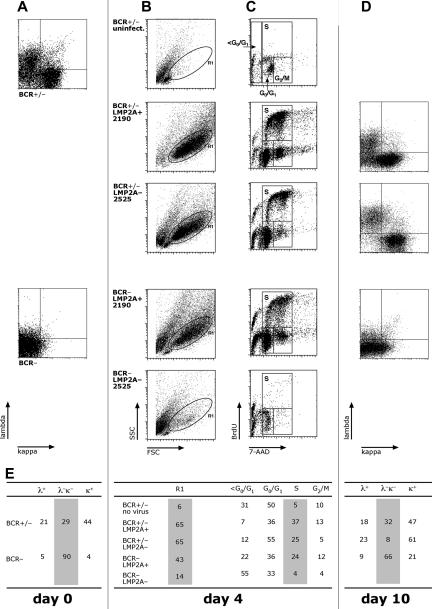
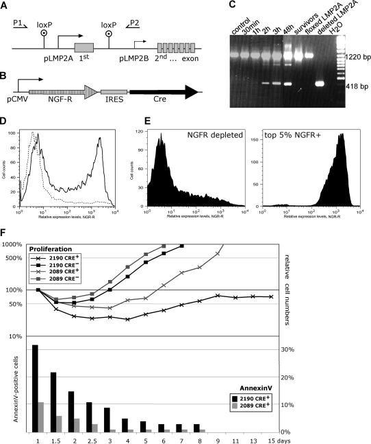
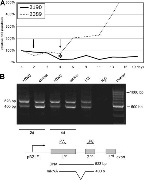
Many cells latently infected with Epstein-Barr virus (EBV), including certain virus-associated tumors, express latent membrane protein 2A (LMP2A), suggesting an important role for this protein in viral latency and oncogenesis. LMP2A mimics B-cell receptor signaling but can also act as a decoy receptor blocking B-cell receptor (BCR) activation. Studies of peripheral B cells have not resolved this apparent contradiction because LMP2A seems to be dispensable for EBV-induced transformation of these B cells in vitro. We show here that LMP2A is essential for growth transformation of germinal center B cells, which do not express the genuine BCR because of deleterious somatic hypermutations in their immunoglobulin genes. BCR-positive (BCR(+)) and BCR-negative (BCR(-)) B cells are readily transformed with a recombinant EBV encoding a conditional, floxed LMP2A allele, but the survival and continued proliferation of both BCR(+) and BCR(-) B cells is strictly dependent on LMP2A. These findings indicate that LMP2A has potent, distinct antiapoptotic and/or transforming characteristics and point to its role as an indispensable BCR mimic in certain B cells from which human B-cell tumors such as Hodgkin lymphoma originate.


Figures

Figure 1
Figure 1
BCR+ but not BCR germinal center B cells infected with LMP2A 2525 EBV enter the S phase of the cell cycle and continue to proliferate in vitro. (A) Primary lymphocytes were isolated from nasal adenoids. The BCR+/− fraction consisted of approximately 30% immunoglobulin surface-negative cells and more than 60% immunoglobulin light chain–positive B cells. The BCR fraction consisted of more than 90% CD19+, light chain–negative B cells. (B) Both fractions were infected with virus stocks of LMP2A+ 2190 EBV or LMP2A 2525 mutant virus and analyzed for forward and sideward scatter characteristics to identify cells in the lymphocyte gate R1 4 days after infection. (C) BCR+/− B cells readily proliferated when infected with either virus (top 2 panels), but cell cycle entry of BCR B cells was observed with LMP2A+ but not with LMP2A mutant EBV (bottom 2 panels). Total cells were analyzed for cell-cycle distribution 4 days after infection by dual parameter flow cytometry with an APC-conjugated α-BrdU antibody to detect incorporation of the nucleotide analog BrdU and with 7-AAD to reveal the cellular DNA content as described. (D) Cell surface expression of λ and κ light chains of cells as in panel A. Ten days after infection, EBV growth-transformed BCR+/− B cells infected with LMP2A+ 2190 EBV (top panel) maintained the typical initial distribution of BCR+ (>60% of λ or κ light chain staining) and BCR (approximately 30%) primary B cells as in panel A. In contrast, BCR B cells were absent 10 days after infection when the same initially heterogeneous BCR+/− fraction was infected with the LMP2A 2525 virus mutant as indicated by the loss of λ- and κ-positive cells in the lower left quadrant (middle panel). The majority of BCR cells infected with LMP2+ 2190 EBV maintained their immunoglobulin surface-negative status (bottom panel). No cells survived when BCR B cells were infected with LMP2A 2525 mutant EBV. (E) Cell-cycle distribution and immunoglobulin light chain surface expression of cells were analyzed.
Figure 2
Figure 2
Sustained LMP2A expression prevents apoptotic death of EBV-transformed B cells. (A) Schematic representation of the LMP2A gene locus in 2190 EBV, which carries 2 loxP sites located upstream of the LMP2A promoter (pLMP2A) and downstream of the first exon of LMP2A. This exon is unique to LMP2A and encodes its amino-terminal signaling domain, whereas the 12 putative transmembrane domains and a short cytoplasmic terminus are encoded by exons 2 through 8, which are shared by the carboxy coterminal LMP2B protein whose transcript is expressed from the LMP2B promoter (pLMP2B). To monitor the status of the LMP2A gene, the PCR primers (P1, P2) were used, which gave rise to PCR products of 1220 bp and 418 bp in length with the parental LMP2A+ 2190 EBV and the Cre-deleted LMP2A variant, respectively. (B) The p3233 expression plasmid is schematically shown, which encodes the truncated NGF receptor (NGF-R) as a phenotypic marker and the site-specific recombinase Cre from a bicistronic transcript. (C) Cre-mediated deletion of LMP2A could be monitored by PCR as early as 2 hours after transfection of p3233. Lanes indicated as floxed LMP2A, deleted LMP2A, and H2O are PCR controls. The lane indicated as “survivors” refers to PCR analysis of 2190-infected B cells (labeled Cre+ in panel F) at 6 weeks after transfection. (D) Transient transfection of the expression plasmid p3233 and FACS analysis of LMP2A+ 2190 EBV LCLs expressing the truncated NGF-R at their cell surface 18 hours after transfection. LCLs established with the prototypic 2089 WT EBV gave comparable results (data not shown). (E) FACS sorting of successfully transfected LMP2A+ 2190 EBV LCLs with the topmost 5% expression levels of NGF-R (Cre+/NGF-R+) versus NGF-R+–depleted (Cre/NGF-R) cells. (F, top panel) The absolute numbers of cells in the different fractions were determined by FACS analysis at indicated days after transfection. BD Biosciences CaliBRITE beads were used as an internal volume standard as described previously. Cells in the lymphocyte gate according to forward and sideward scatter criteria were included in the analysis and set to 100% 24 hours after transfection. EBV-infected LCLs infected with LMP2A+ 2190 EBV carrying the floxed LMP2A allele are indicated (2190, black lines) as well as LCLs infected with the prototypic 2089 WT EBV strain (2089, gray lines). Transfected and sorted Cre+/NGF-R+ cell fractions are indicated (Cre+) and compared with cells depleted from NGF-R+ cells by sorting (Cre). (Bottom panel) Cre-transfected (Cre+/NGF-R+) LCLs infected with either 2089 WT EBV or LMP2A+ 2190 EBV were analyzed for annexinV staining by FACS at the time points indicated.
Figure 3
Figure 3
Continuous proliferation of EBV-transformed cells requires LMP2A. (A) The absolute numbers of B cells infected with 2089 WT EBV or 2190 EBV with its LMP2A gene flanked by 2 loxP sites were determined by FACS analysis as in Figure 2F. The cells were transduced with the E coli–derived Cre protein HTNC for 3 hours, and the surviving cells were counted by FACS and set to 100% on day 1. 2089 WT EBV–infected LCLs recovered from this manipulation and started to proliferate, but LCLs infected with LMP2A+ 2190 EBV did not recover within the observation period. The asterisk indicates the time point when cells were removed for quantitative real-time PCR analysis of the LMP2A gene, which revealed that more than 90% of the 2190 EBV–infected cells had lost the LMP2A allele 4 days after transduction (data not shown). (B) Reverse transcription (RT)-PCR analysis of BZLF1 transcripts in HTNC-transduced 2190-infected LCLs 2 and 4 days after Cre transduction. The positions of the primer oligonucleotides P7 and P8 are schematically shown together with the predicted size of the PCR products indicative of mRNA-specific cDNA molecules of the actively transcribed BZLF1 gene and (contaminating) viral DNA. BZLF1 transcription levels did not increase but showed a slight reduction upon deletion of LMP2A, indicating that its loss did not trigger the onset of the lytic phase of EBV's life cycle.

References

    1. Vol 70. Lyon, France: IARC; 1997. Epstein-Barr virus and Kaposi's sarcoma herpesvirus/human herpesvirus 8: International Agency for Research on Cancer. Anonymous.
    1. Rickinson AB, Kieff E. Epstein-Barr virus. In: Knipe DM, Howley PM, Griffin DE, et al., editors. Fields Virology. 5th ed. Vol 2. Philadelphia, PA: Lippincott Williams & Wilkins; 2007. pp. 2655–2700.
    1. Kieff E, Rickinson AB. Epstein-Barr virus and its replication. In: Knipe DM, Howley PM, Griffin DE, et al., editors. Fields Virology. 5th ed. Vol 2. Philadelphia, PA: Lippincott Williams & Wilkins; 2007. pp. 2603–2654.
    1. Bell AI, Groves K, Kelly GL, et al. Analysis of Epstein-Barr virus latent gene expression in endemic Burkitt's lymphoma and nasopharyngeal carcinoma tumour cells by using quantitative real-time PCR assays. J Gen Virol. 2006;87:2885–2890. - PubMed
    1. Altmann M, Pich D, Ruiss R, Wang J, Sugden B, Hammerschmidt W. Transcriptional activation by EBV nuclear antigen 1 is essential for the expression of EBV's transforming genes. Proc Natl Acad Sci U S A. 2006;103:14188–14193. - PMC - PubMed

Publication types

MeSH terms

Substances